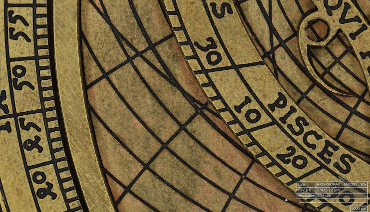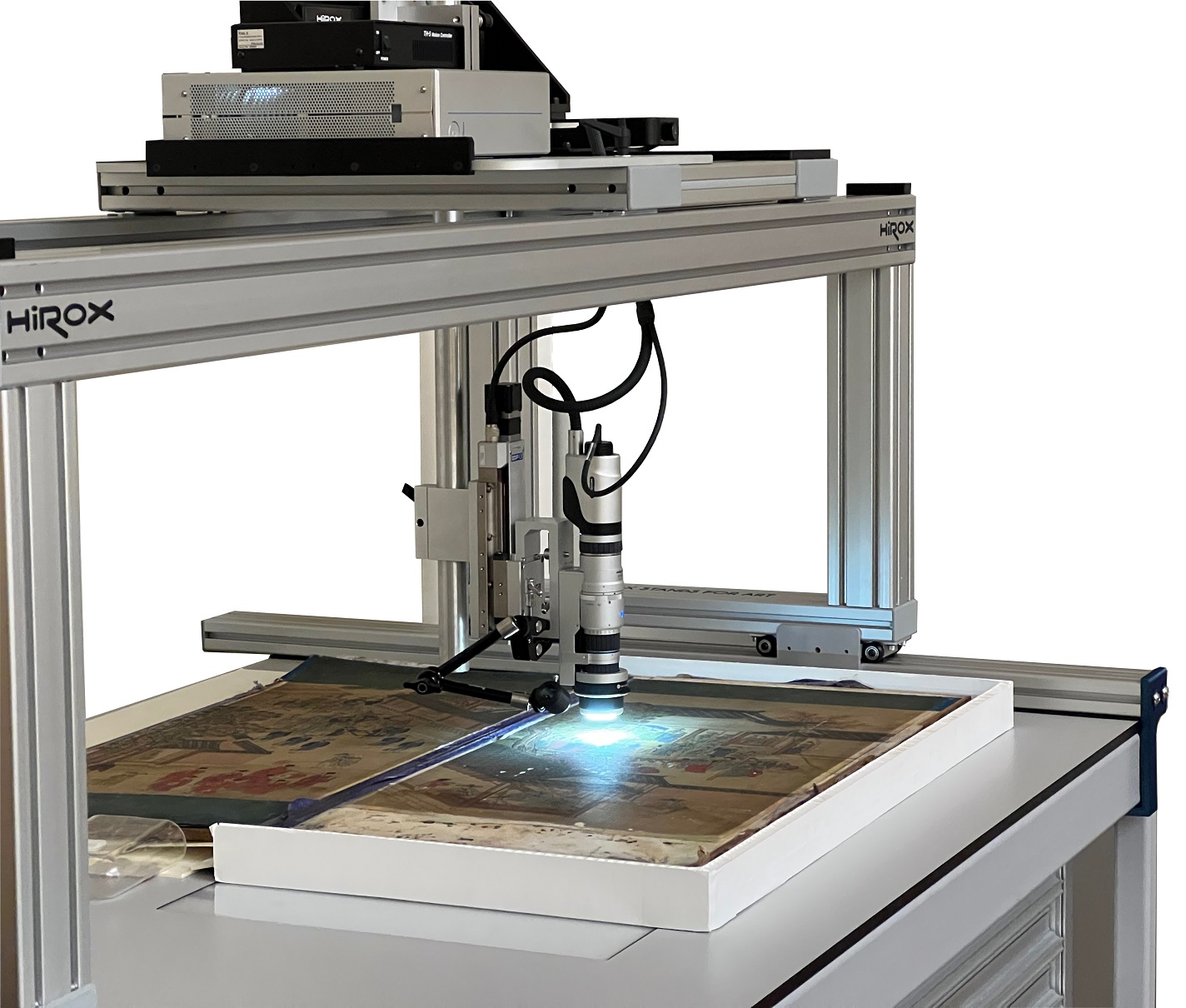
40+ years of innovation in quality control Exclusive Hirox partner Demos with your samples
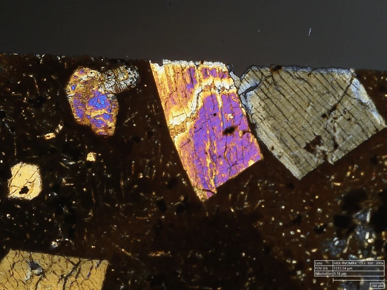
Simitecno IT Book a demo
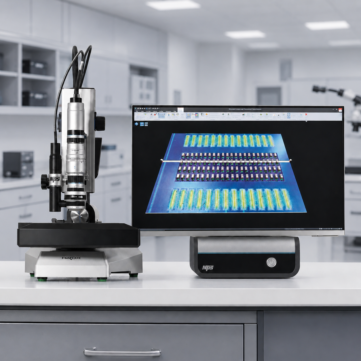
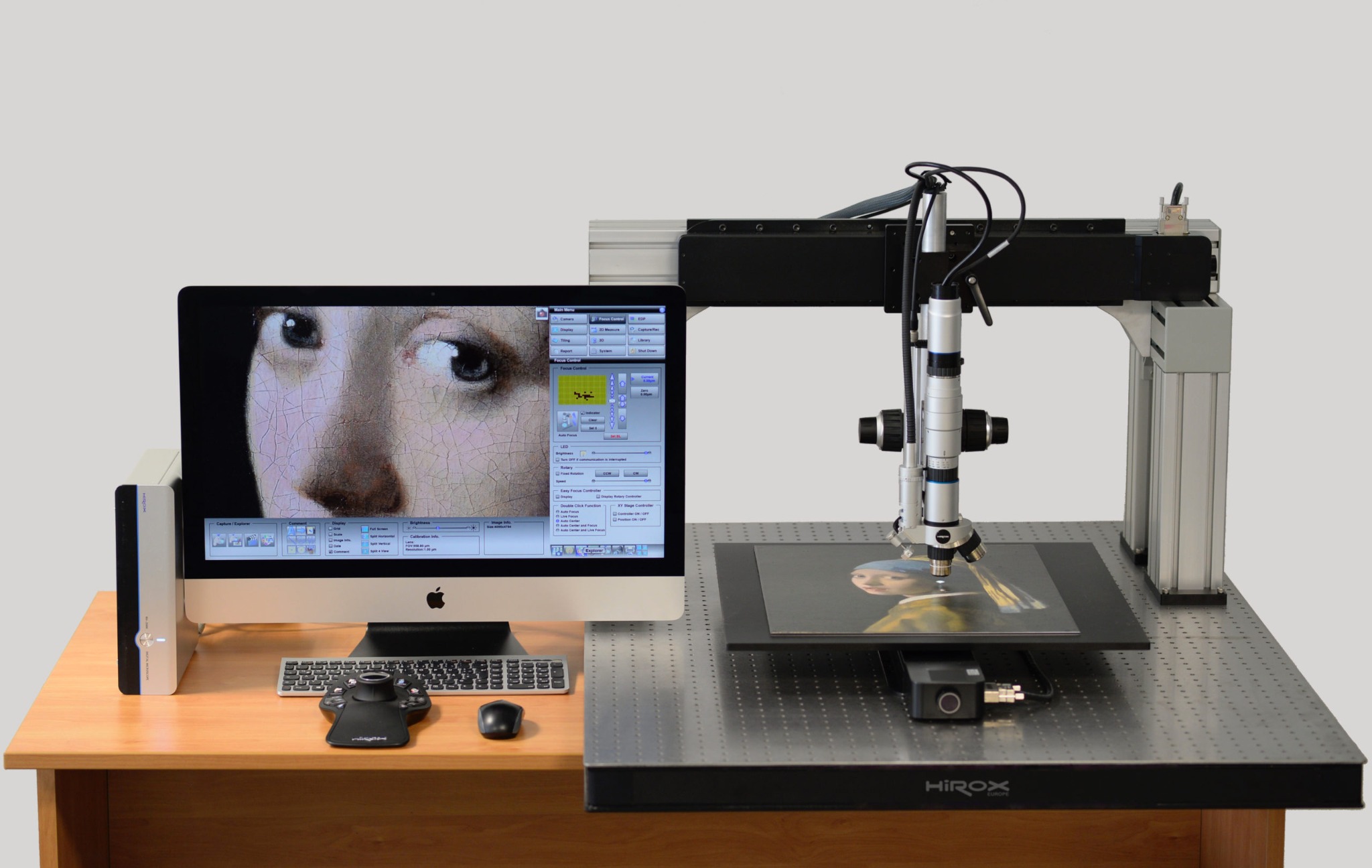
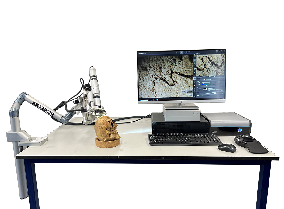
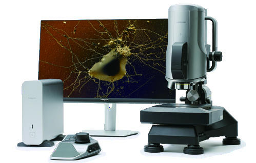
HIROX HRX-02 digital microscope with monitor and controller

INDUSTRY · RESEARCH · QUALITY CONTROL

See the defects that compromise quality.

3D digital microscopy, advanced metrology and automation for reliable inspection decisions.

Simitecno video

FROM THE PROBLEM TO THE MEASUREMENT

We do not start from the instrument.
We start from your application.

Describe the component, material or defect to be analysed. Simitecno specialists identify the most suitable technology and arrange an application trial on your own sample.

TECHNOLOGIES

A complete platform to understand and control materials

See all products →
NPS confocal profilometer
Confocal profilometry

NPS

Non-contact scanning of profiles, surfaces and roughness with nanometre resolution.

NPS features →
Simitecno Demo Centre at IIT Milan

DEMO CENTRE @ IIT MILAN

Try the technology on a real case

The Demo Centre at the Center for Nano Science and Technology of the Istituto Italiano di Tecnologia is dedicated to 3D digital microscopy and advanced optical metrology.

Book a demonstration

TRADE FAIRS AND WORKSHOPS · CALENDAR AND ROADMAP

Next events and workshops

In program and in preparation: explore the technologies, applications and test opportunities on your samples.
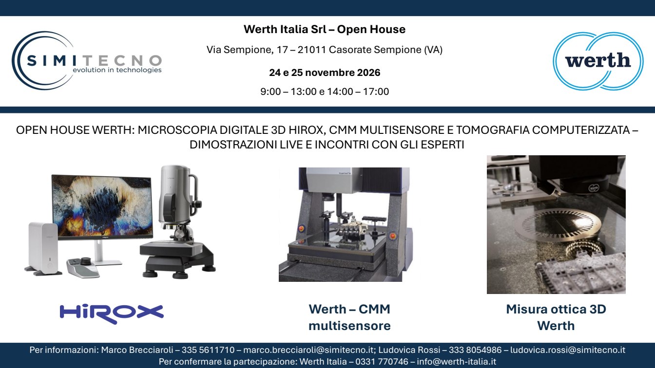
Werth Open House flyer

OPEN HOUSE

24 and 25 November 2026

Open House Werth Italia

Casorate Sempione (VA)

Live demonstrations of Hirox 3D digital microscopy, multisensor CMMs and computed tomography.
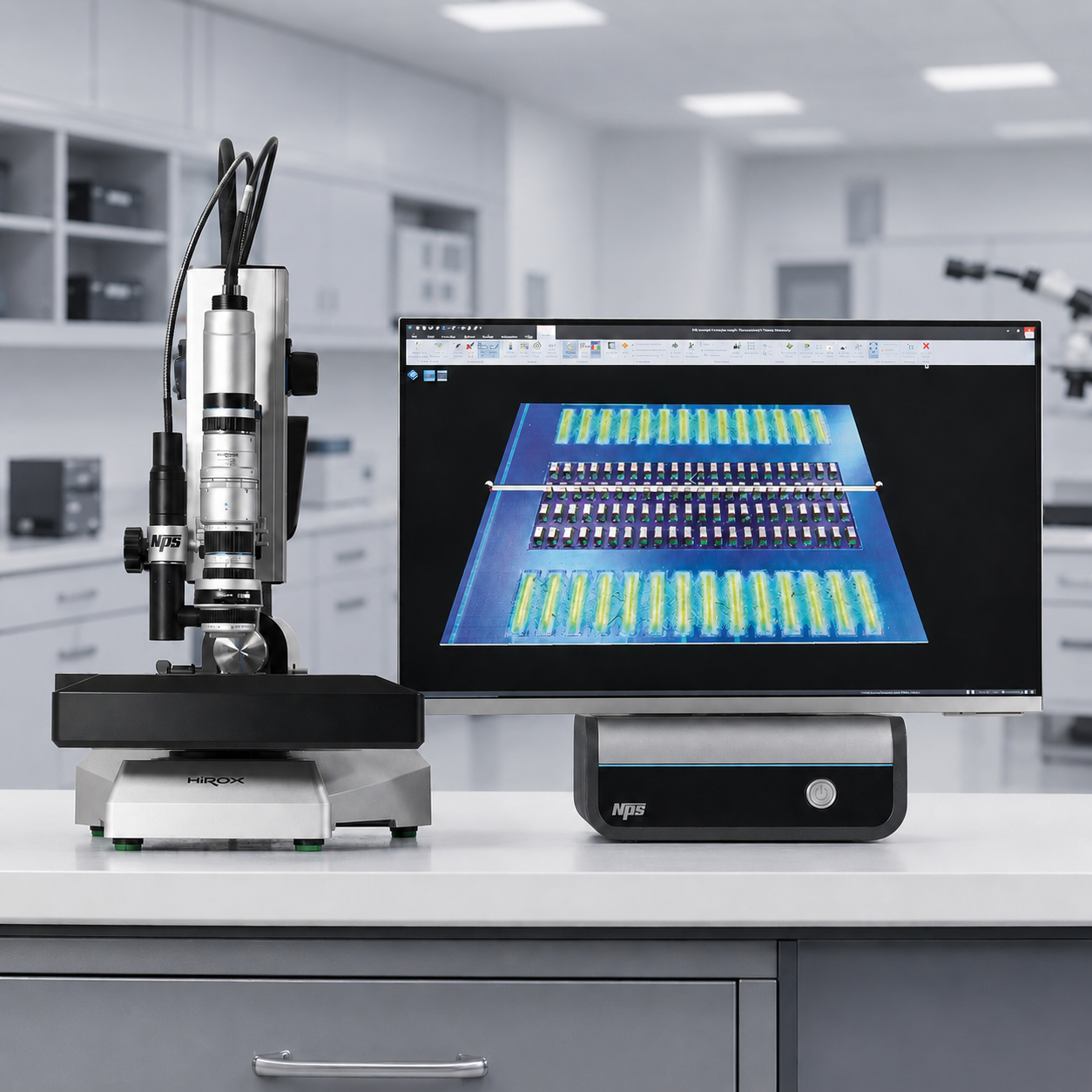
System for the IIT Workshop – Milan

WORKSHOP

Dates to be confirmed

IIT workshop – Milan

IIT · CNST · Milan

Automated microdefect detection and 3D surface topography

3D digital microscopy, structured-light scanning and AI for in-line quality control.
Exposed instrumentation

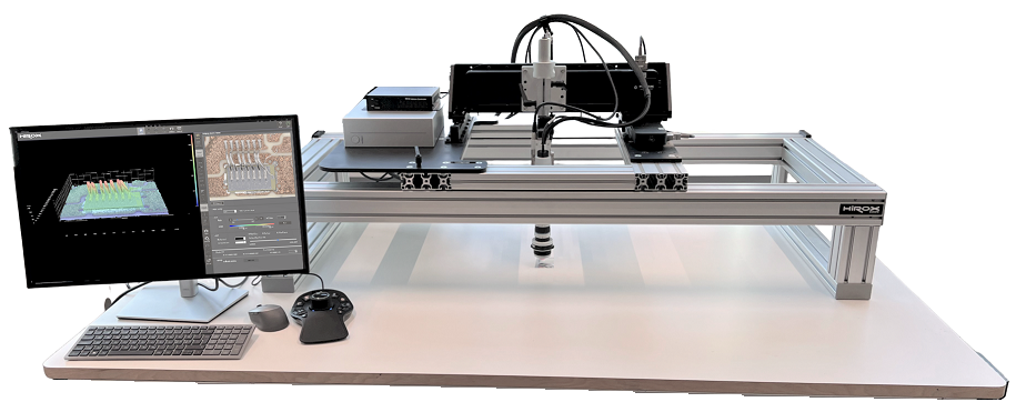
  • Hirox with benchtop stand
  • NPS

THEMATIC TRACKS

Upcoming workshop roadmap

Seventeen application tracks dedicated to materials analysis. Dates and venues are being finalised.
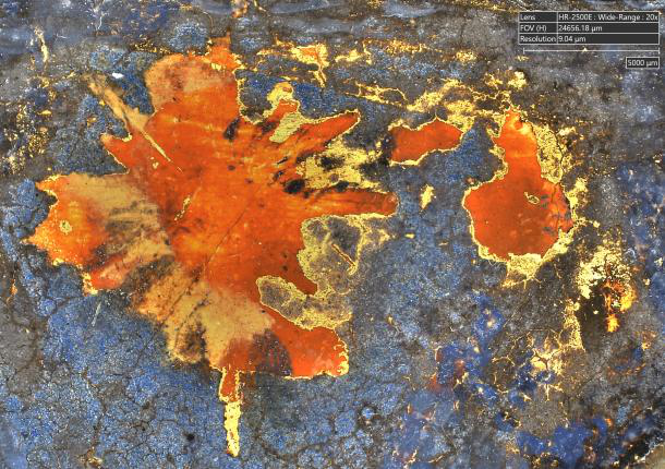
Registration and demos on request by e-mail: we agree attendance and samples together.

2026 · 2025

Past events

See the events in program →
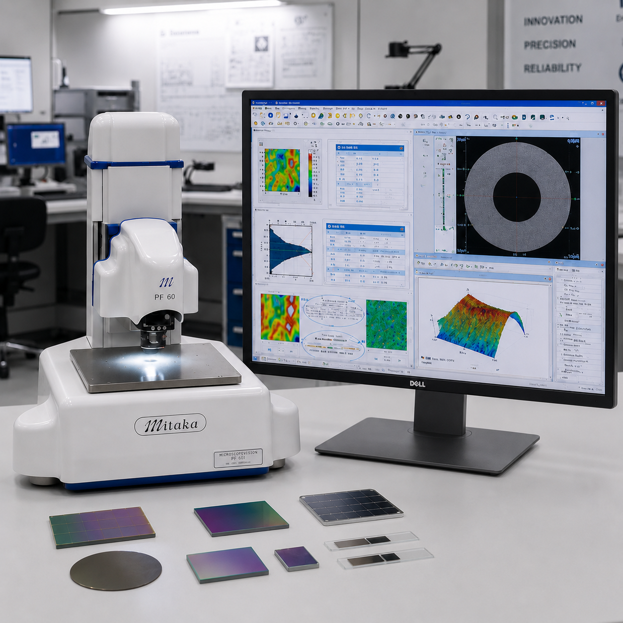
System for the CNR ISM Frascati Workshop

WORKSHOP

09 October 2026

CNR ISM workshop – Frascati

Frascati

Announced instrumentation

  • 1 Mitaka
  • 1 Hirox with Rvomax + NPS
System for BI-MU – Milan

TRADE FAIR · LIVE TESTING

13–16 October 2026

BI-MU – Milan

Fieramilano Rho · Hall 22 · Stand E31

Announced instrumentation

  • 1 gantry with Hirox
  • 2 Hirox with benchtop stands, one of them with NPS
  • 1 cobot with Taraz
  • 1 cobot cell with Hirox
System for A&T – Vicenza

TRADE FAIR · APPLICATION DEMOS

27–29 October 2026

A&T – Vicenza

Vicenza

Announced instrumentation

  • 2 Hirox with benchtop stands, one of them with NPS
  • 1 cobot with Taraz
MECSPE · Bologna

ARCHIVE · 2025

5–7 March 2025

MECSPE · Bologna

Industrial fair dedicated to technologies for manufacturing and quality control. Simitecno presented its solutions and collected application contacts.

Workshop CNR ISAC · Bologna

ARCHIVE · 2025

11 April 2025

Workshop CNR ISAC · Bologna

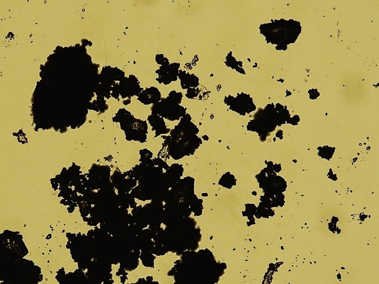
Application meeting on particulate matter and cultural heritage, with 3D digital microscope and surface analysis.

Macro–Micro–Nano is Politecnico di Milano

ARCHIVE · 2025

17–19 June 2025

Macro–Micro–Nano is Politecnico di Milano

Three days of multi-scale demonstrations with HIROX HRX‐02, NPS and Polytec Micro.View.

NanoInnovation · Rome

ARCHIVE · 2025

2025

NanoInnovation · Rome

Presentation and demonstrations dedicated to the characterization of nanostructured materials and surfaces.

ESB · Turin

ARCHIVE · 2025

7–11 September 2025

ESB · Turin

Participation in the conference on biomaterials: 3D microscopy and optical metrology for the research and control of surfaces.

SurfaceLab · Rome

ARCHIVE · 2025

2025

SurfaceLab · Rome

Application meetings on surfaces and biomaterials, with comparison of imaging and measurement needs.

Content 2025 summarizes the known initiatives; photographs, presentations and additional reports will be integrated when available.

VIDEOS AND DEMONSTRATIONS

Simitecno Italia YouTube channel

The channel collects application videos, demonstrations and presentations of the quality control technologies offered by Simitecno and its partners: 3D digital and confocal microscopy, profilometry, optical metrology, 3D scanning, tomography, automation and inspection robotics.

Visit the channel and watch the videos

DO YOU HAVE A DEFECT TO IDENTIFY?

Show us your sample.
We will show you how to measure it.

Start the analysis

SIMITECNO INSTRUMENTS

Comprehensive materials inspection, from 3D surface analysis to internal structure and chemical composition.

Explore images, features, applications and configurations. Each card leads to the full technical page and to a sample trial request.

3D DIGITAL MICROSCOPY

Observation, measurement and automation

Modular systems to analyse details, surfaces and complex components in the laboratory or in production processes.

Hirox HRX-02 3D digital microscope3D microscopy

Hirox 3D Digital Microscope

High-resolution three-dimensional scanning and measurement, 360° observation and magnification up to 10,000×.

  • Multifocus and 2D/3D stitching
  • Metrology and particle analysis
  • Motorised rotary optics
Go to the instrument →
NPS confocal profilometerConfocal profilometry

NPS profilometer

White-light chromatic confocal profilometer for non-contact scanning of profiles and surfaces.

  • Resolution down to 76 nm in Z
  • Acquisition up to 2,000 points/s
  • Roughness, form and topography
Go to the instrument →
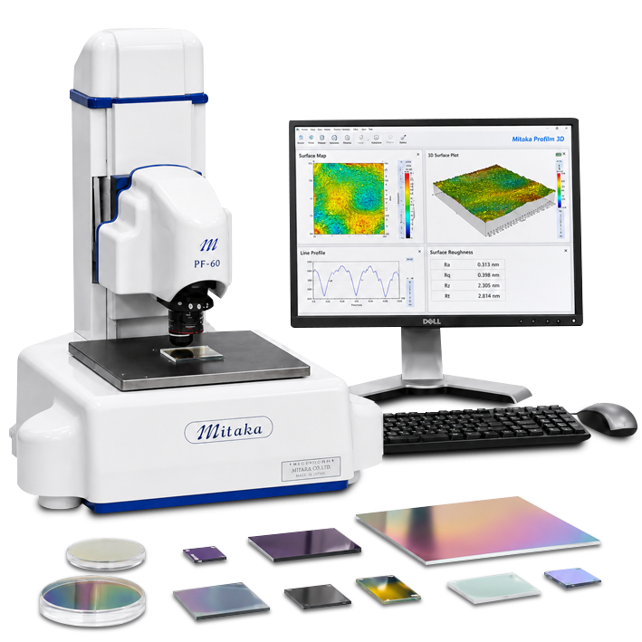
Laser profiler Mitaka PF-60 on a laboratory benchLaser metrology

Mitaka PF‑60 laser profilometer

Real-time 3D measurement with single-point focusing, non-contact and without field stitching.
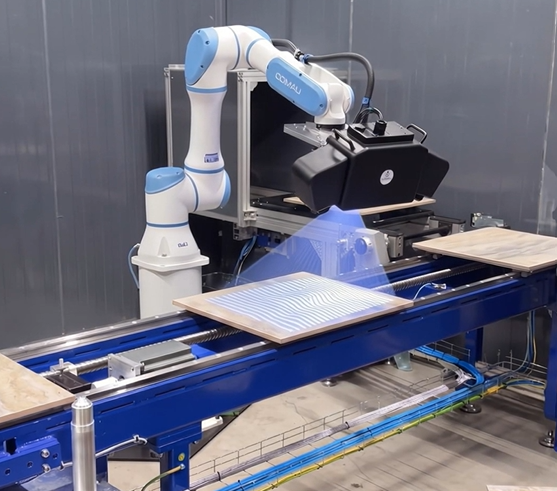
  • Autofocus scale resolution 0.01 µm
  • X × Y × Z range: 60 × 60 × 60 mm
  • Opaque, transparent and reflective surfaces
Go to the instrument →
Taraz ASM system on COMAU robots with fringe projection on a panel

Taraz ASM

Blue-light fringe projection optical system to measure surface topography, form and roughness without contact.

  • Four fields of view and resolutions available
  • Acquisition in 2 seconds or less
  • ISO 25178 analysis with TarazSurf
Go to the instrument →
HiroxBot Station with digital microscope and collaborative robot COMAURobotics and AI

HiroxBot

The integration of HIROX with collaborative robots automates inspection, quality control and measurement.
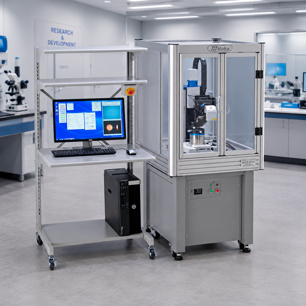
  • Repeatable recipes and positions
  • PCB and component inspection
  • Reduced operator variability
Go to the instrument →
Five-axis Mitaka MLP-3PAll-around scanning

5-axis Mitaka laser profilometer

Laser CMM to measure forms, profiles and internal and external surfaces from different directions.
  • 5 programmable motorised axes
  • Components up to 120 mm
  • Steep angles below 87°
Go to the instrument →
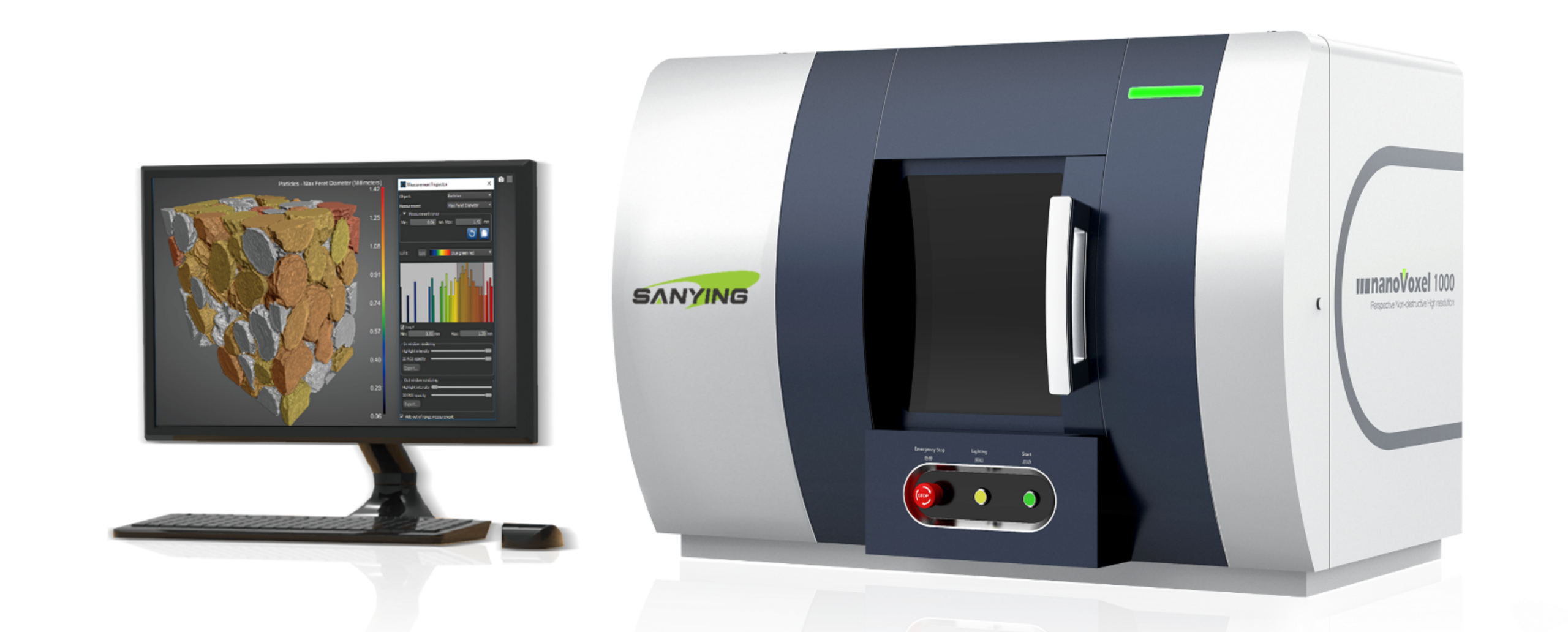
nanoVoxel-1000 benchtop CT scannerX-ray tomography

Benchtop CT scanner

Compact desktop micro‑CT system to observe defects, porosity and internal structures without sectioning the sample.
  • Detectability down to 300 nm
  • Sealed 110 kV source
  • Optional in-situ 4D imaging
Go to the instrument →
Light fringe projection for post-process metrological controlPost-process inspection

Taraz PM

Optical station for complete 3D measurement of complex components using fringe projection, photogrammetry and multi-view acquisition.

  • 3D reconstruction from multiple viewpoints
  • Comparison against CAD reference and tolerances
  • Final inspection in the workshop and in production
Go to the instrument →

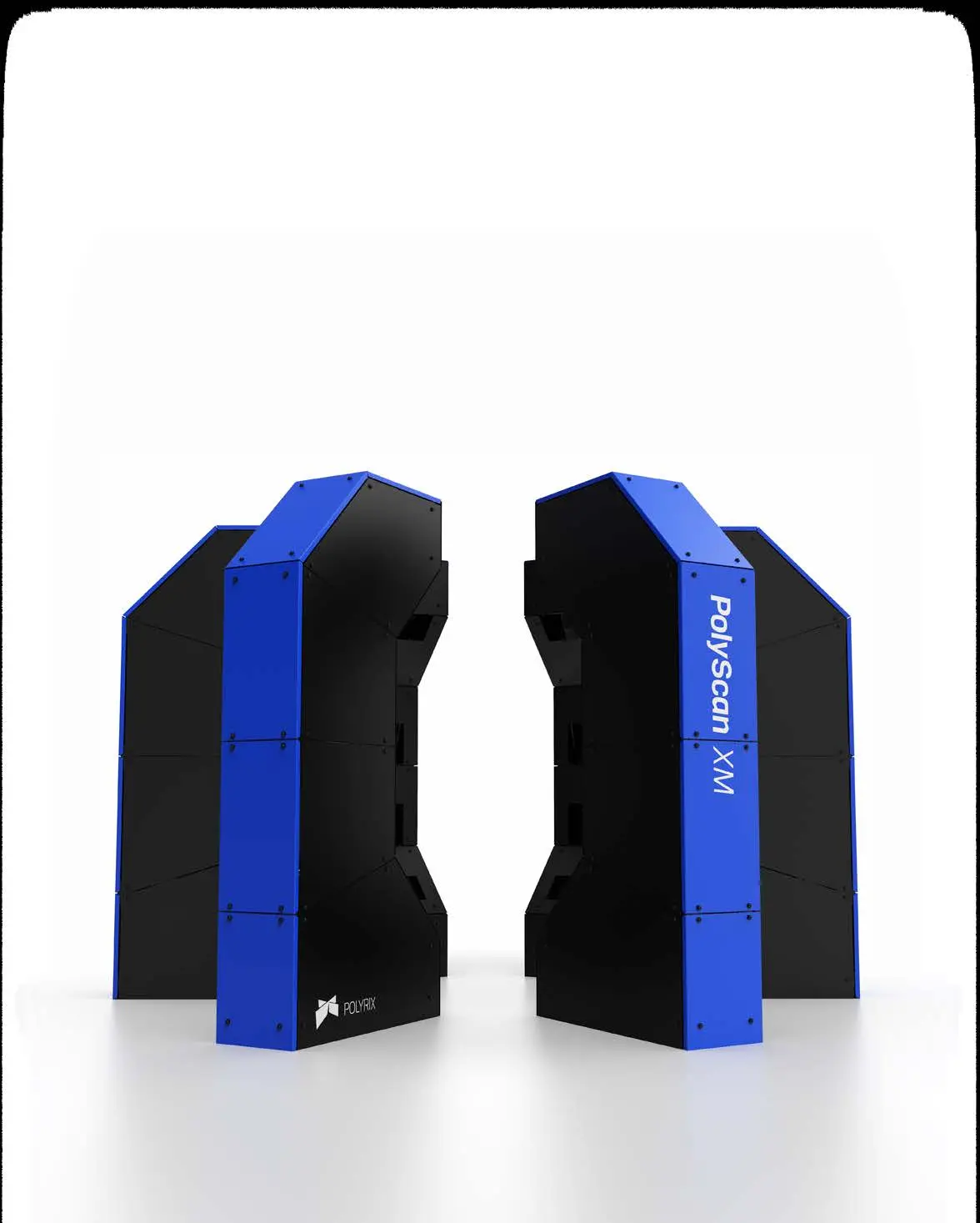
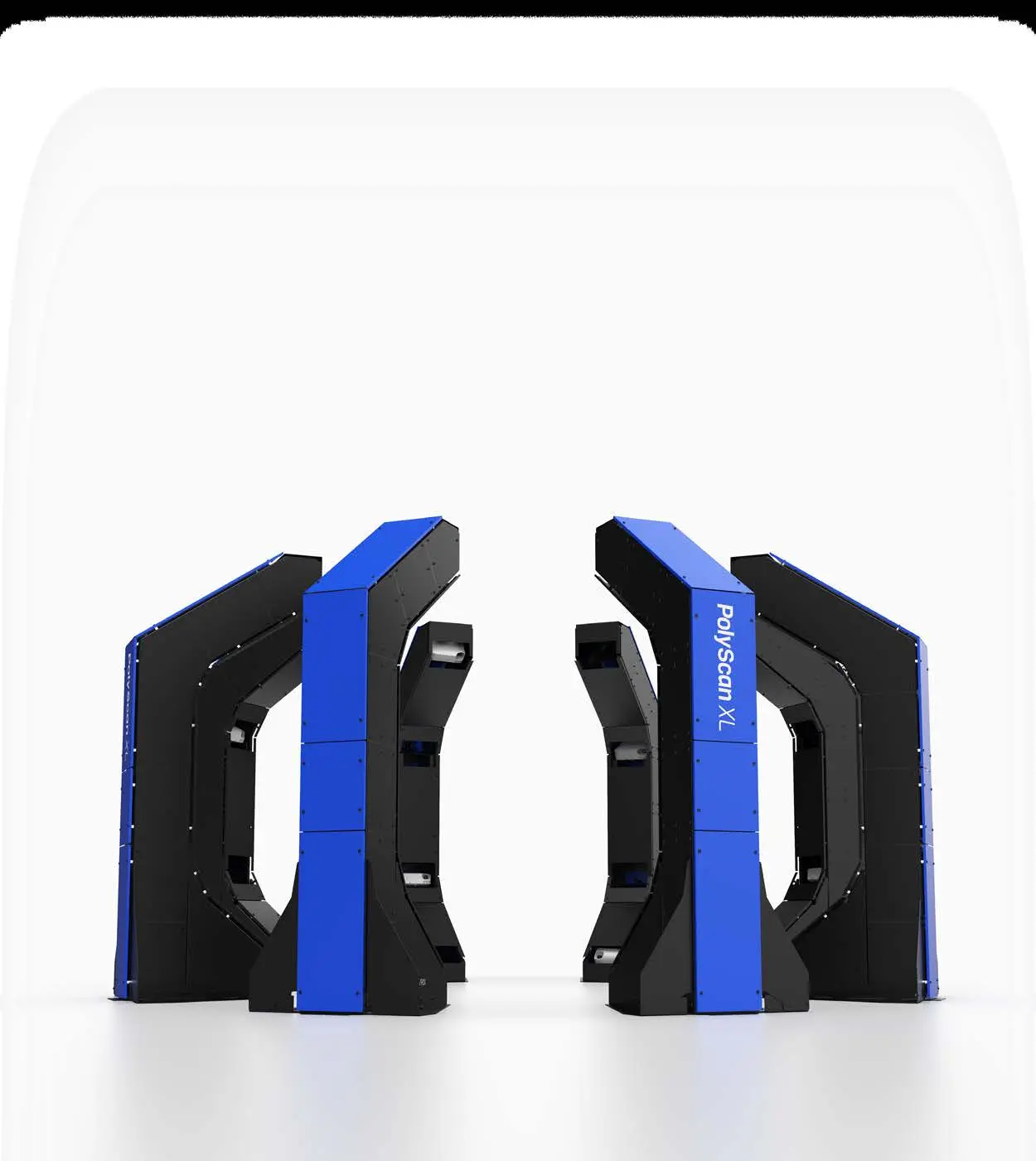
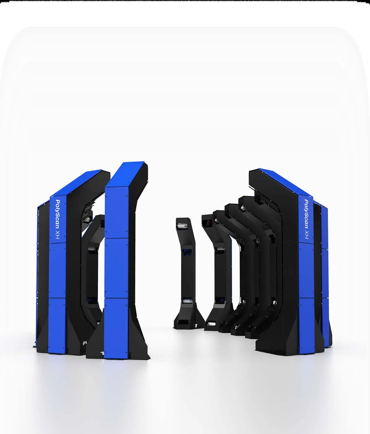
POLYRIX · 3D SURROUND METROLOGY

PolyScan XS, XM, XL and XH

Multi-sensor systems with no moving parts, capturing the entire component in a single cycle, from small details to structures of 6.5 metres and beyond.

Discover the PolyScan family

CAN’T FIND THE RIGHT CONFIGURATION?

Describe your sample and the result you need to achieve.

Ask a specialist

APPLICATIONS

Find the right solution for your industry

Each application links the problem, the measurement method, the result and the recommended technology.

02

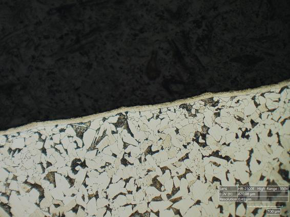
Metallography

Microstructures, inclusions, fractures, corrosion, roughness and failure analysis.

Assess the material →
03

Aerospace and automotive

Complex components, critical surfaces, reverse engineering and automated inspection.

Study the inspection →
07

Pharmaceutical industry

Tablet coating, particle size, particle count, roughness and batch-to-batch control.

Explore the application →
09

Surface fabrication

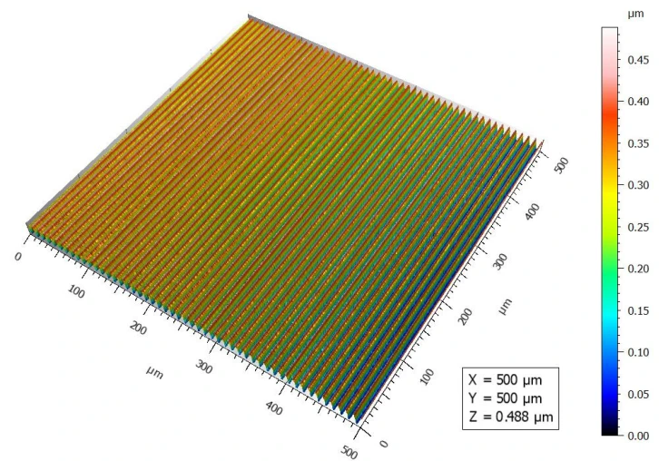
3D surface weaving map measured with Mitaka PF-60

Surface weaving range evaluation with Mitaka PF-60 and comparison of results with and without S filter.

Open the publication (PDF) →
← Back to applications

HIROX FOR MICROELETTRONICA

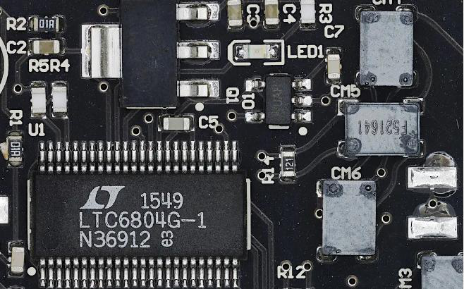
3D inspection of circuits, packages and microcomponents

From electronic assembly to failure analysis: high-resolution observation, 360° inspection, 2D/3D measurements and contactless confocal profilemetry.

Analyzes a component
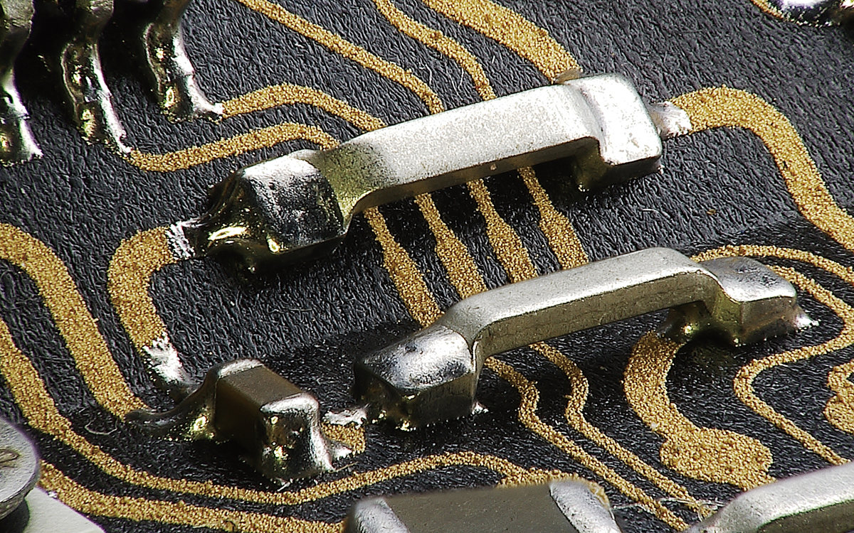
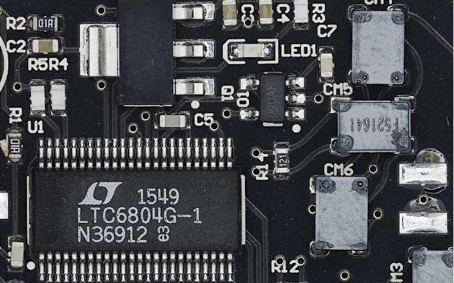
High resolution detail of bonding and tracks of a microelectronic circuit

HIROXBOT · ELECTRONIC

Automated optical inspection of electronic cards

Digital and robotic microscopy to observe components, tracks and welds and document controls on PCB cards.
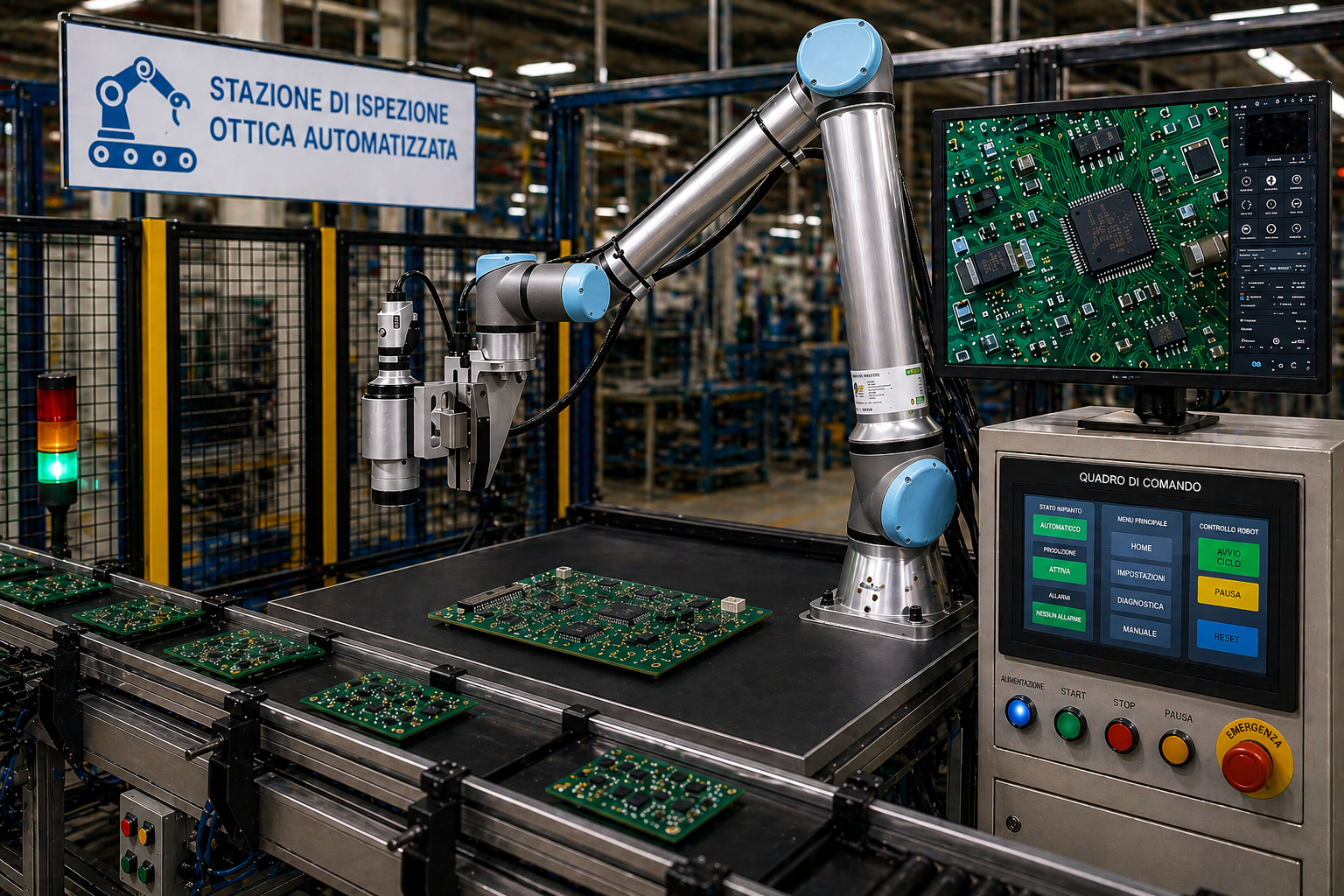
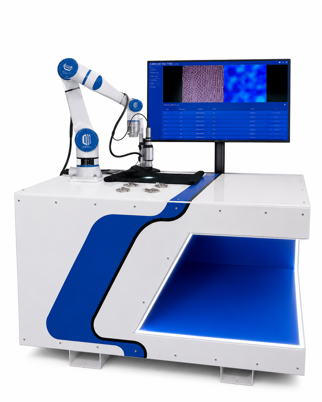
HIROXBOT: Robotized station for optical inspection of PCB electronic cards
HIROXBOT — Automated optical inspection station for electronic applications.

AUTOMATIC OTTICAL METHOD

Automatic measurement of PCB microsections

From calibrated images of the HIROX digital microscope, the software measures copper layers, dielectric thicknesses, metallic walls and microway filling. It works offline and generates annotated images, PDF reports and CSV files for each sample.

PCB Microsections with annotated measurements and comparison between software values and manual measurements

DISTINTIVE FUNCTIONS

See the defect, measure it and document it

The configuration is adapted to circuit, package, wafer, connector or microcomponent.

360°

Rotating optics

Inspection of pins, welds and geometries hidden by multiple angles without manipulating the component.

LIVES

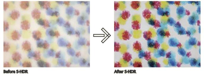
Focus and HDR

Full-focus images and reflex control on metals, resins and glossy surfaces.

2D/3D

Automatic Tiling

High-resolution and continuous 3D reconstructions of circuits and extended areas.

μm

Quantitative measures

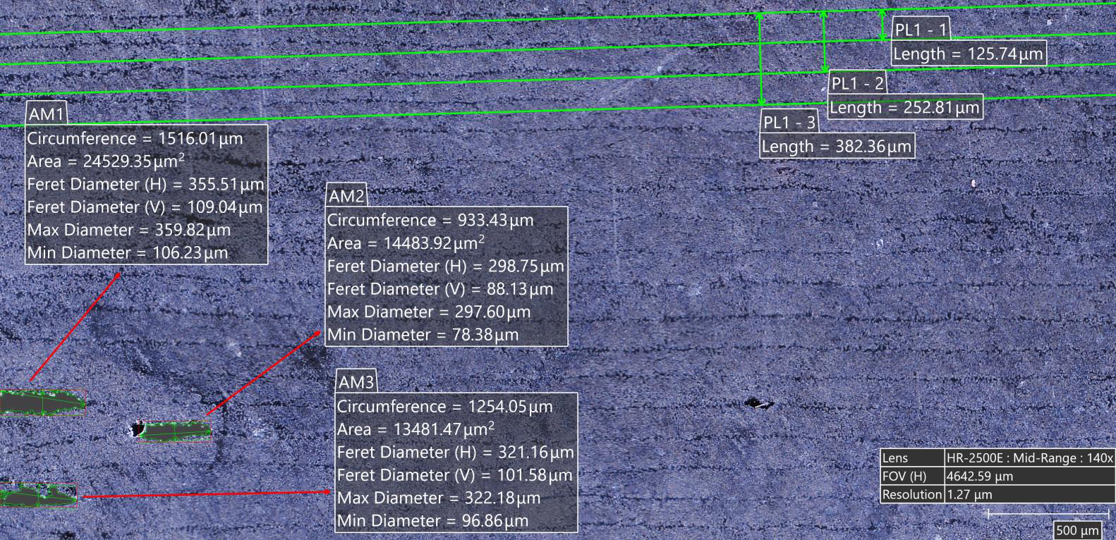
Lengths, areas, diameters, angles, heights, volumes, profiles and roughness Ra/Rz.

IR - UV

Selective illumination

Coaxial, annular, diffuse, polarized, IR and UV for different materials and contrasts.

LIVE FOCUS

More focal planes, a single sharp image

Combine focal planes to obtain a focus image even on samples with height variations.
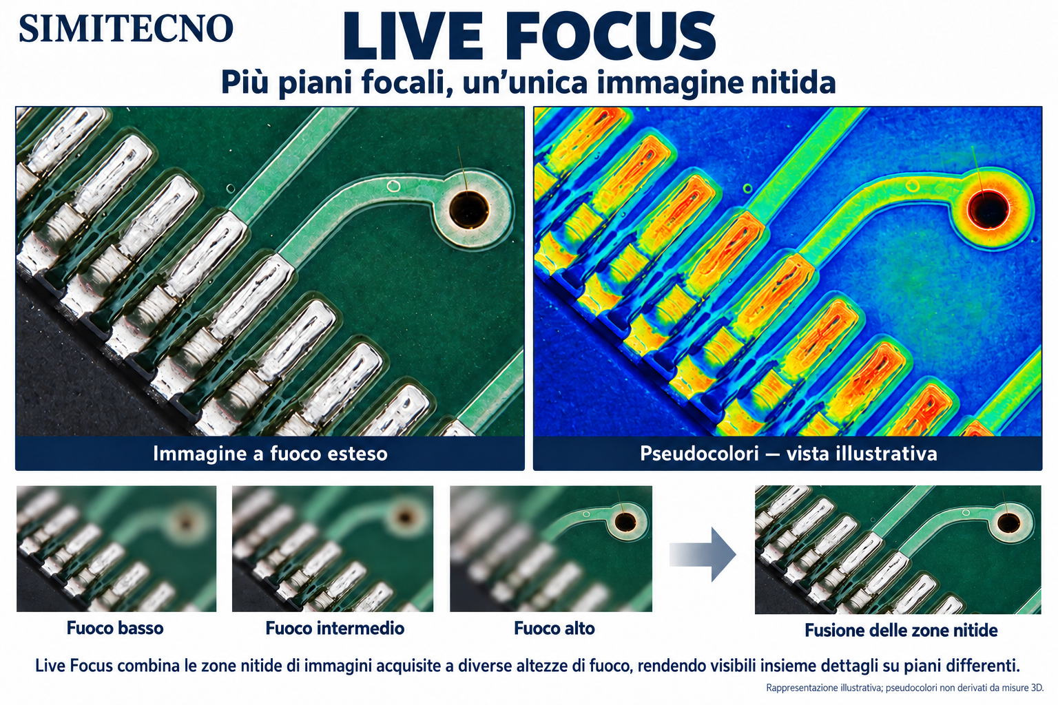
SIMITECNO Live Focus: welded contacts on PCB, pseudo-colored explanatory view and pattern of fusion of the sharp zones of different focal planes
Live Focus — fusion of the nitid zones acquired on different focal planes. Illustrative image; pseudocolors do not result from 3D measures.

APPLICATION SCHEDE

Controls along the entire electronic process

Electronic Assembly Inspection
01 · ASSEMBLY

Welding, BGA and bonding

Welded joint control, mounted components, bonding defects and flux residues.
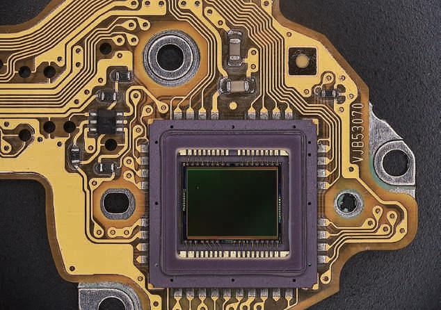
Integrated circuit observed with HIROX
02 · MICROCHIP

Reverse engineering

Observation of patterns, pistes, transistor and intra-chip details for study and comparison.
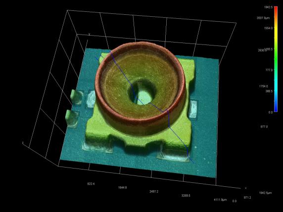
3D reconstruction of a microcomponent
03 · METROLOGY

2D/3D dimensional measurements

Profiles, heights, volumes, roughness and pseudo-color patterns, wireframe or real color.
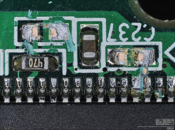
Electronic card for high resolution mapping
04 · MAPTURE

Stitching of extended circuits

Automatic union of 3D images and scans through coordinated movements XY and Z.
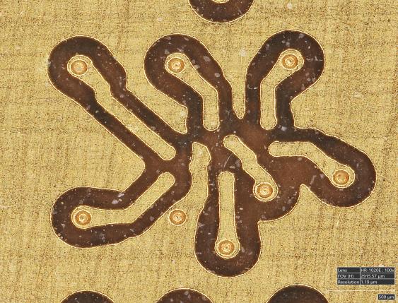
Detail of bonding and electronic tracks
05 · FAILURE ANALYSIS

Crimes and process defects

Search for microcracks, fractures, delaminations, short circuits and interrupts of the tracks.
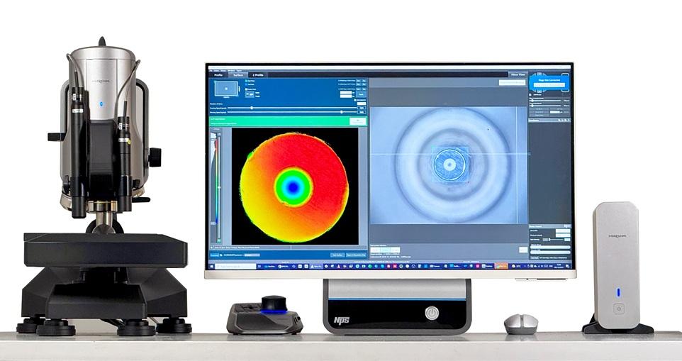
HIROX system with NPS confocal sensor
06 · NPS

Profiles and confocal surfaces

Planarity, copulity, wave, texture, defects and roughness on complex surfaces.
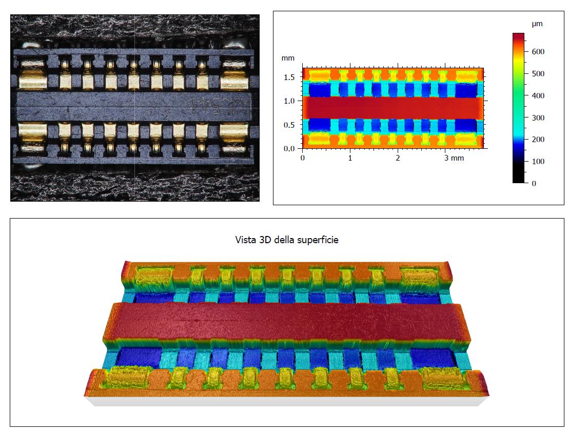
A COMPONENT TO ANALYSISTE?

We define optics, lighting and measurements on the sample.

Request a trial
Electronic assembly
ASSEMBLY AND PACKAGING

Control of joints without prior separation

Rotating optics, coaxial light and multifocus highlight shape, wetability, residuals and welding anomalies even on difficult geometries.

Microchip
REVERSE ENGINEERING

From the intra-chip retail package

Guide Map, motorized zoom and coordinates allow you to correlate the general vision with patterns, pistes and microscopic structures.

3D measurement
MEASURES 2D/3D

Geometry and morphology in a single environment

Measures of length, area, height, volume and roughness are performed directly on 3D images and models.

Extended circuit
AUTO TILING

Continuous documentation of cards and wafers

The XY automatic movement combines adjacent frames; Z scan adds three-dimensional topography without discontinuity.

Electronic Difference
ANALYSIS FAILURE

Locate the origin of the failure

Selective illuminations and angular observation help to distinguish cracks, fractures, delaminations and interrupts of the tracks.

NPS sensor
CONFOCAL PROFILED

Measure exactly what you see

The NPS sensor acquires up to 2,000 height information per second and generates contactless 3D profiles and surfaces.

← Back to applications

HIROX FOR FARMACEUTICAL INDUSTRY

3D inspection of tablets, coatings and granules

A correlative platform to verify uniformity of the coating, morphology, roughness, size, particles and variations between batches.

Analyze a sample
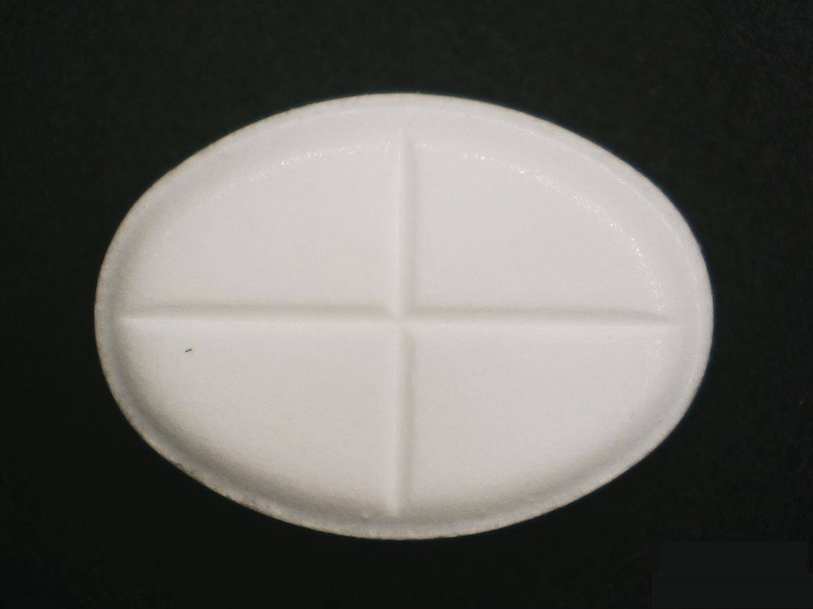
Pharmaceutical tablet analyzed with HIROX

CONTROL FARMACEUTICAL QUALITY

From the whole tablet to the microstructure

HRX-RVOMAX, 3DT software and NPS sensor integrate high magnification imaging and surface metrology.

30×

Macro Inspection

General documentation of the tablet, engravings, edges and dimensional characteristics.

1500×

Coating Analysis

Evaluation of the uniformity of the film by coaxial light and focus variation.

3D

Topography and reliefs

Reconstruction of the surface, size of letters, carvings, heights and volumes.

COUNT

Granulate analysis

Counting and classification of particles by shape and size.

2D

dimensional control

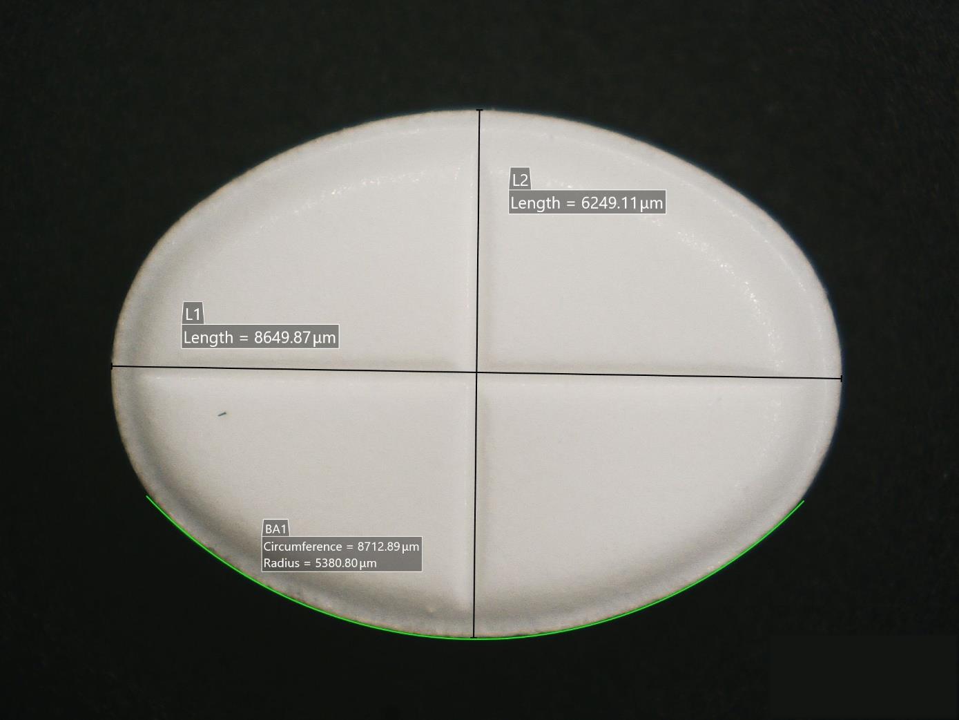
Diameter, arc, area, perimeter and geometries of the whole or sectional tablet.

60 nm

Rugour NPS

Interoperable confocal profilemetry with HIROX and MountainsMap metrological reports.

APPLICATION SCHEDE
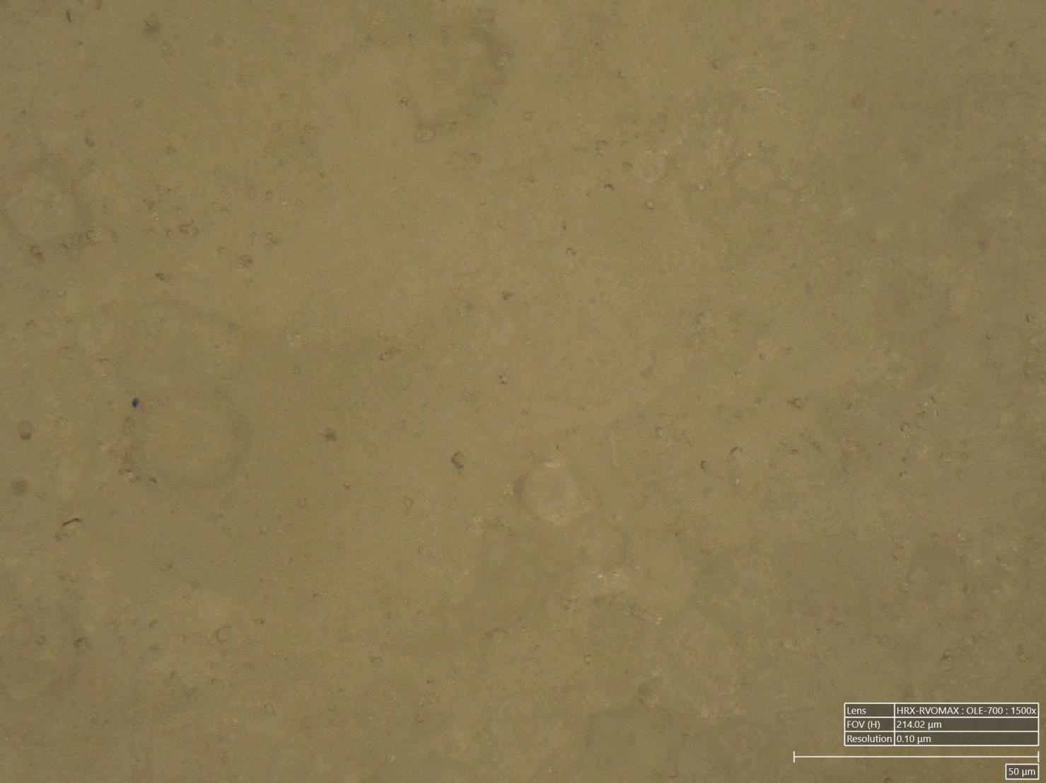
Analysis performed on the pharmaceutical sample

High Enlargement Coating Surface
01 - COATING

Uniformity of coating

1500× analysis with coaxial lighting and 3D reconstruction of the surface.
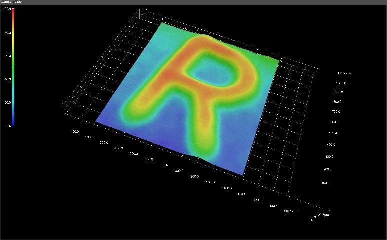
Embossed letter reconstructed in 3D
02 · RELIVE

Size of letters and markings

Height, profile and morphology of embossed details with 3D model export.
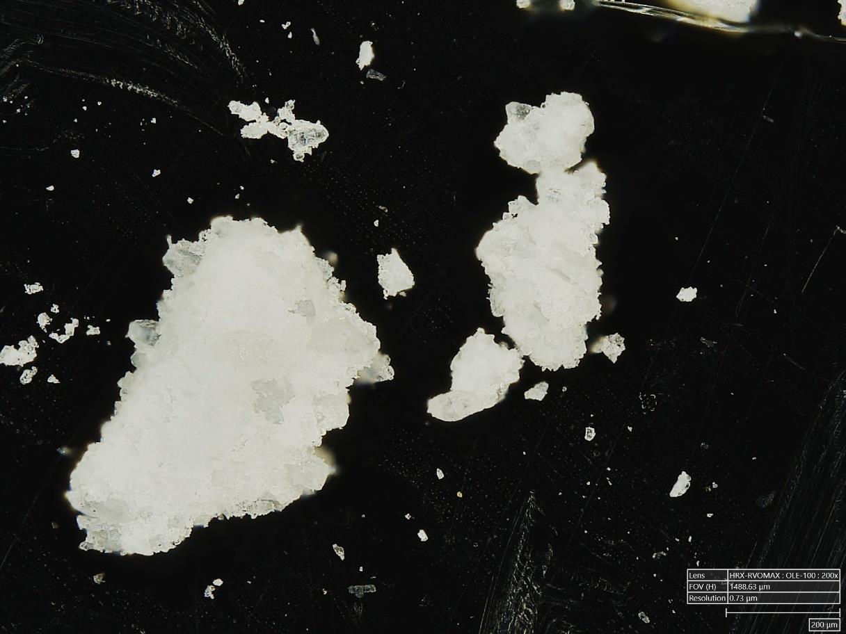
Granulated of the tablet in reflected light
03 · GRANULATION

Reflected and transmitted light

Comparison of granulate morphology on matt support and on glass.
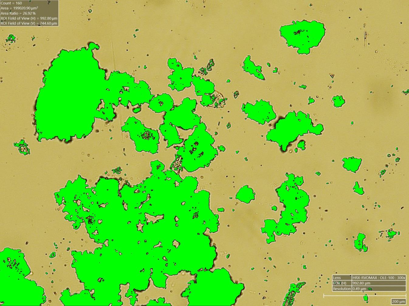
Automatic particle count
04 · PARTICULAR

Particle Count and Auto Area

Counting, classification and dimensional parameters with CSV and Excel export.
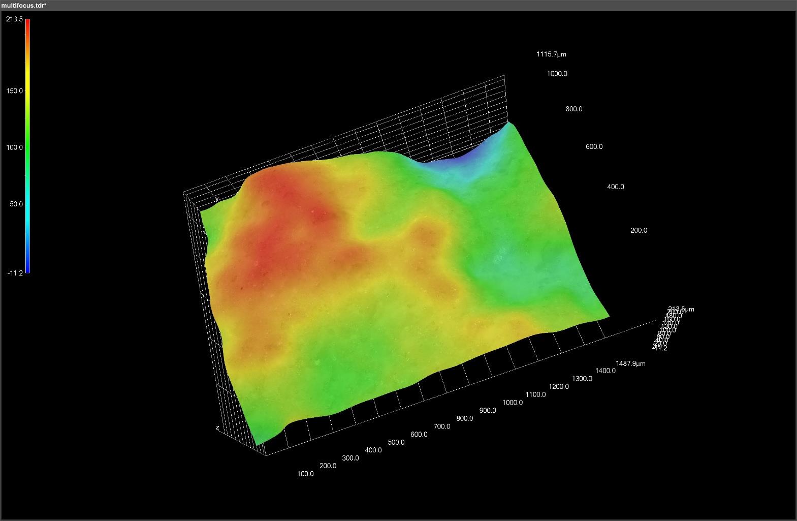
3D Topography of the carved section
05 · SECTION

Tablets carved

Observation of the interior, roughness of the carving and three-dimensional reconstruction.
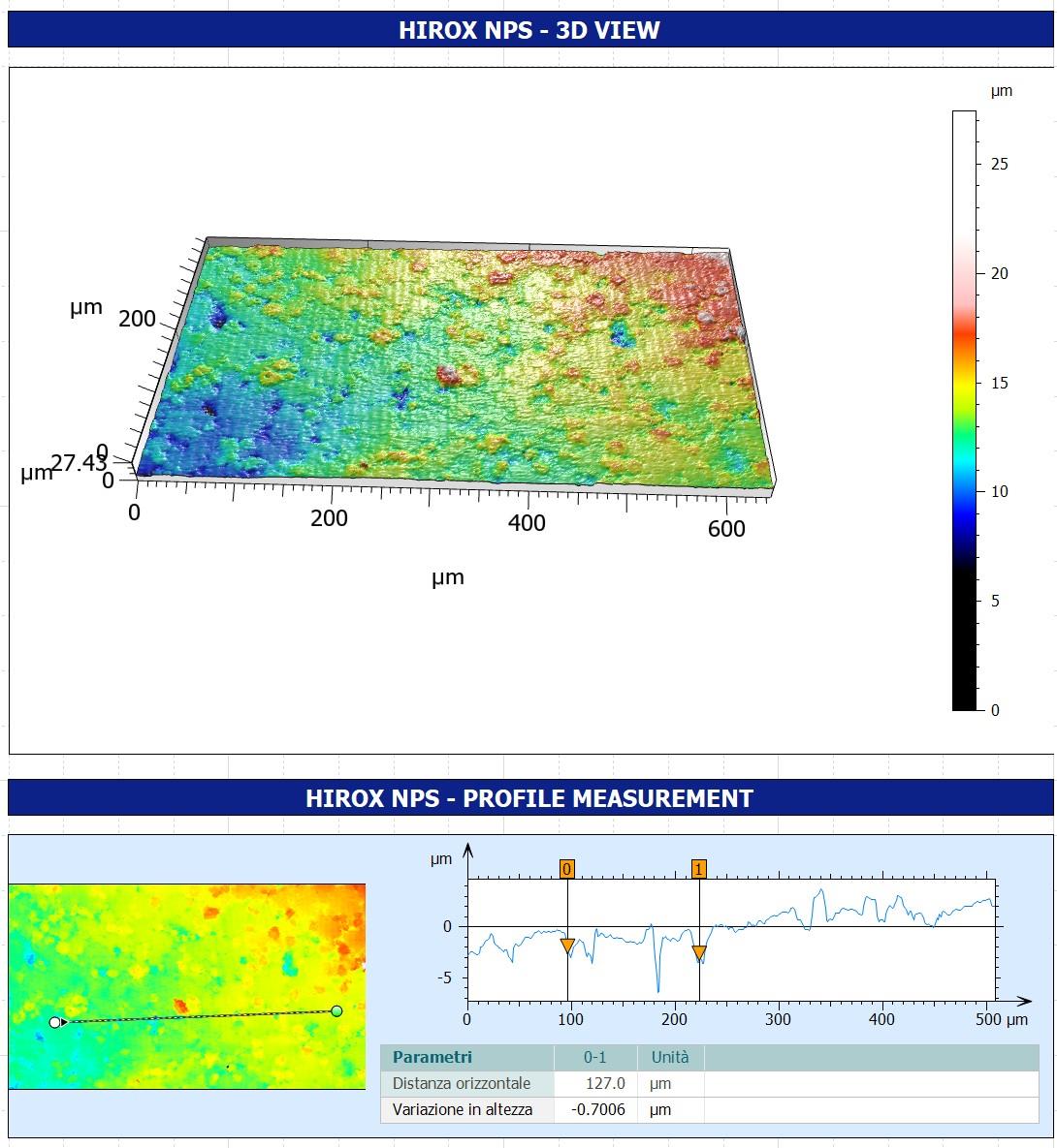
Surface metrological report
06 · NPS

ISO compliant wheels

Confocal profile with Z resolution of 60 nm and MountainsMap reporting.
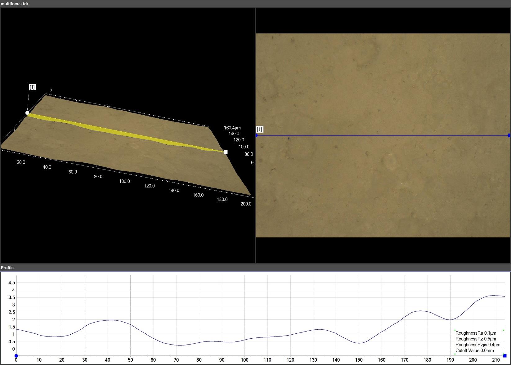
A CONFRONTAL LIGHT?

We set up a repeatable protocol of imaging and measurement.

Request a trial
Coating
REVIEW

Uniformity and microdefects of the film

Coaxial light and focus variation generate images and topographies to compare homogeneity, texture and coating defects.

Relief
3D RICOSTRATION

Height and shape of markings

The 3D model allows you to extract profiles and measure the height of letters and symbols; data can also be exported as a cloud of points.

Granulated
GRANULATION

Two illuminations to read morphology

The reflected light highlights the surface on opaque support; the transmitted light shows contours and distribution on slide.

Particle count
COUNT PARTICULAR

Automatic counting and classification

The particles are separated by brightness and color and classified by area, perimeter, diameters and shape; the results feed CSV/Excel reports.

Section 3D
COMPRESSA INTAGLIA

Internal structure and roughness of cutting

The 200× analysis documents the interior of the tablet and produces 3D profiles and reconstructions of the section.

NPS
CONFOCTION SESSION

Observed surface metrology

The Z resolution of 60 nm, the integration with HIROX and MountainsMap allow repeatable measurements and reports according to ISO parameters.

← Back to applications

HIROX + MITAKA FOR SOTTILI FILM

3D imaging and nanoscale surface measurement

An integrated solution to document the film, locate defects and discontinuity, reconstruct topography and measure contactless thicknesses and steps.
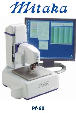
Laser profiler Mitaka PF-60 for thin film measurement
HIROXImaging 2D/3D
MITAKA PF-60Profile and step height

CORRELATIONAL METHOD

From optical location to quantitative measurement

The two technologies work on scales and complementary objectives: HIROX identifies and documents the area; PF-60 measures the profile with autofocus laser probe.

01 · HIROX HRX-02

3D Exploration and Documentation

  • Live Focus for surfaces with altitude variations.
  • Live HDR for reflective films and critical contrast zones.
  • Guide Map to find the region of interest.
  • Car 2D/3D Tiling for extensive reconstructions.
  • Polarized lighting, coaxial, oblique and annular.
  • 2D/3D measurements, roughness and automatic reports.
02 · MITAKA PF-60

High resolution laser profile

  • Laser spot probe from 0.5 μm.
  • XYZ standard area: 60 × 60 × 10 mm.
  • XY Resolution: 0.1 μm.
  • Resolution Z: 0.01 μm / 0.001 μm.
  • Direct measurement of heights, edges and steep corners.
  • Automatic scanning of wide and diversified surfaces.

FUNCTIONS FOR SOTTILI FILM

Controls from the visual defect to the nanometer step

LIVES

Extended focus

Images fully on fire even on edges, steps and surfaces not perfectly planar.

HDR

Reflex management

Real-time visualization of clear and dark areas on reflective coatings and substrates.

MAP

Navigation of the ROI

The image at low magnification becomes a map connected to the position of the XY table.

2D/3D

Automatic Tiling

Continuous mosaics and topographic maps of larger areas of the individual field of view.

Ra · Rz

Profile and roughness

Profile curves, ripple and roughness parameters on selected lines or areas.

DATA

Measures and reports

Images, capture parameters, coordinates, results and transferable statistics in reports.

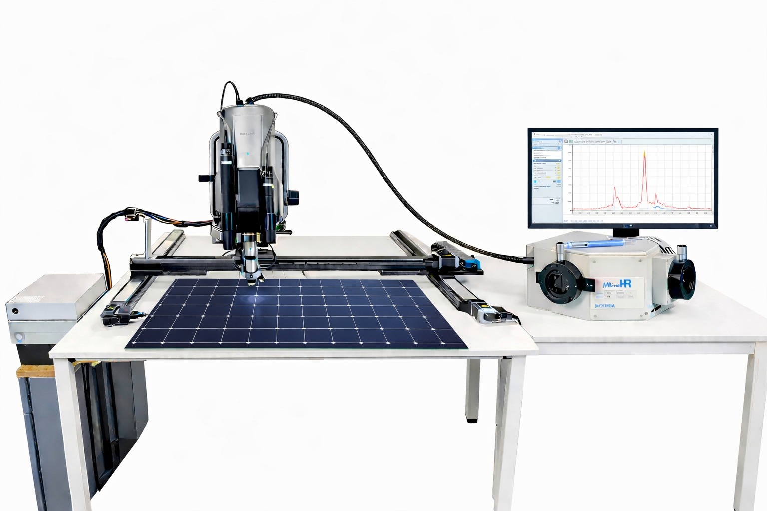
APPLICATIONAL CASES · FOTOVOLTARY PROVISIONS

Nb₂O₅ niobium oxide film on borosilicate glass

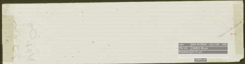
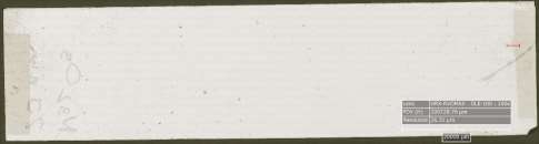
The illustrated sample presents a film deposited by PVD evaporator with a nominal thickness of 20 nm. The analysis combines the optical reconstruction of the entire film with the measurement of the step between substrate and coating.

Nominal thickness
20 nm
Profile distance
2,005μm
Variation in height
-19,95 nm
2D reconstruction of thin film with polarized light
2D reconstruction with polarized light
2D reconstruction of thin film with coaxial light
2D reconstruction with coaxial light

OTTENABLE RESULTS

A complete application card of the film

Thickness and step height

Quantitative confirmation of the thickness deposited along selected profiles.

Uniformity and topography

Distribution of local quotas and variations on the analyzed area.

Surface defects

Pin-hole, particles, edges, scratches, discontinuity and contamination.

Traceability

Coordinates, images, parameters and measures comparable to samples and processes.

A FILM TO CHANGE?

We verify imaging and measure directly on the sample.

We define area, substrate, finish, waited step and required resolution.

Request a trial
← Back to applications

HIROX FOR CONSERVATION

3D microscopy for cultural heritage

Non-invasive scientific documentation, pre/post comparison restoration and quantitative analysis of surfaces, stratigraphy and fragile materials.
DISTINTIVE FUNCTIONS

A single platform, from reperto to measure

Modular optics and statics allow to work on small fragments, paintings, fabrics, manuscripts, sculptures and non-moving objects.

360°

Rotating optics

Observation from multiple angles without turning or manipulating the reperto.

LIVES

Multifocus and HDR

Completely on fire and reflex control even on irregular surfaces.

2D/3D

Automatic Tiling

High resolution mosaics and continuous 3D reconstructions of extended areas.

UV · IR

Special lighting

Polarized, oblique, diffused, UV and IR light to make readable hidden details.

μm

Quantitative measures

Profiles, heights, areas, roughness and geometric comparison between analysis campaigns.

DATA

Traceability and reporting

Metadata, coordinates, images and measurements organized in repeatable reports.

APPLICATION SCHEDE

Six areas of investigation

Select an application to see objectives, HIROX configuration, specific functions and observation examples.

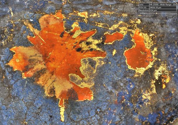
Paint surface with pigments and craquelure
01 · DIPINTI

Painted surfaces

Craquelure, lifts, touches, abrasions and comparison before/after restoration.
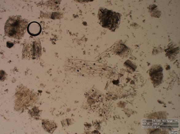
stratigraphic section observed under the microscope
02 · CROSS-SECTION

Materials and stratigraphy

Thicknesses, interfaces, pigment distribution and sequence of application layers.
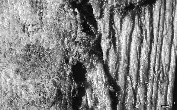
microscopic detail of historical fabric
03 · TEXTS

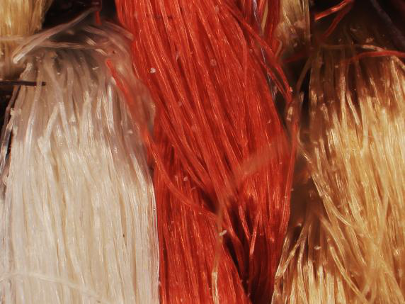
Fabrics and fibres

Ingots, yarns, abrasions, contaminants and non-invasive assessment of the conservative state.
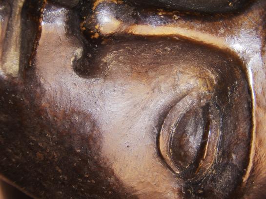
Lapidea surface detail
04 · 3D SUPERFICI

Sculpture and stone materials

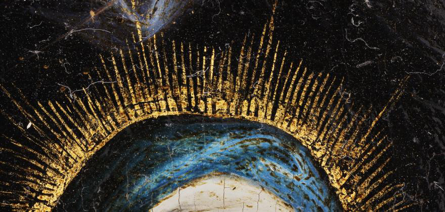
Microfessure, erosion, incrustations, roughness and volumetric loss.

Historical manuscript observed at high resolution
05 · CAR AND PERGAMENA

Manuscripts and miniatures

Traits, gildings, pigments, engravings, annotations and alterations of fragile supports.
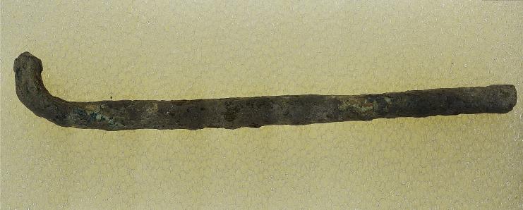
Archaeological metal reperto
06 · REPERTIES

Archaeology and metals

Patins, corrosion, processing traces, encrustations and restoration monitoring.

A DOCUMENTAL REPERT?

We build the configuration around the work.

We value geometry, fragility, area to be acquired, lighting and measures required.

Request a trial
High resolution pictorial detail
PITTORICAL SUPERFICI

Document, compare and measure without contact

The combination of large depth of field and 3D topography allows to describe craquelure, waxing, lifting, abrasions and deformations of the pictorial film.

Main surveys

  • High-resolution mapping of damaged areas.
  • Comparison of pre/post restoration work.
  • 3D reconstruction of deformations and dislivelli.
  • Texture analysis to distinguish original and reintegration.

Dedicated HIROX functions

  • Car 2D/3D Tiling for extended areas.
  • Live Focus for non-planar surfaces.
  • Bright, polarized and HDR light for pigments and reflections.
  • Guide Map to find the same area of interest.
Layer sample in section
MATERIALS AND STRATIGRAPHS

Read the strata sequence

The observation of glossy cross-section allows to distinguish preparation, pigment, paint, interfaces and discontinuity with repeatable measurements of the thickness.

Main surveys

  • Measure of the thicknesses of the individual layers.
  • Study of overlapping and color transitions.
  • Detection of irregular gaps and interfaces.
  • Identification of mineral particles and aggregates.

Dedicated HIROX functions

  • Automatic 2D measurements of distance, area and thickness.
  • Motorized zoom and memorized coordinates.
  • Coaxial lighting, diffused and polarized.
  • Auto Report for comparable documentation with Raman or SEM-EDS.
Trama of historical fabric
ALL AND FIBRE

Observe braids and degradation without compressing the artifact

The high working distance and modular staves allow to analyze fragile fabrics, embroidery and extended surfaces while keeping the sample free.

Main surveys

  • Weaving techniques, weaving and embroidery techniques.
  • Morphology of natural and synthetic fibres
  • Wear, tears, abrasions and loss of flexibility.
  • Deposits, powders, molds and surface contaminants.

Dedicated HIROX functions

  • Live Focus for yarn at different odds.
  • Directional light and radiant for reliefs and dislivelli.
  • Tiling 2D to document extended portions.
  • Time Lapse and coordinates for time monitoring.
Surface of stone material
SCULTURE AND MATERIALS LAPIDEI

Quantify erosion, roughness and material loss

The 3D reconstruction makes cracks, microfessures, abrasions, intersections and morphological variations on complex surfaces.

Main surveys

  • Microfessure, cracks and discontinuity.
  • Erosion and atmospheric or anthropogenic wear.
  • Organic deposits and saline encrustations.
  • Comparison of the original surface, consolidated and reintegrated.

Dedicated HIROX functions

  • 3D profile, height differences and volumetric loss.
  • Ra/Rz roughness on selected areas.
  • 360° rotary optics for reliefs and subsquadri.
  • Bridge, T-Stand and arm for non-moving works.
Detail of manuscript
MANOSCRITTI, MINIATURES AND CARTA

Reveal contactless details

The combination of high-resolution imaging and selectable illuminations highlights strokes, engravings, doratures, pigments, restorations and alterations on paper and parchment.

Main surveys

  • Calligraphic traits, dry engravings and doratures.
  • Pigments on paper and parchment.
  • Folds, scratches, abrasions and tarlo holes.
  • Annotations, repentances and faded details.

Dedicated HIROX functions

  • UV, IR, polarization and oblique light.
  • HDR in real time for clear and dark areas.
  • Optical macro for complete work and micro for detail.
  • Imaging and uncontacted measurement on fragile supports.
Archaeological finding
ARCHEOLOGY AND METAL

Potatoes, corrosion and traces of processing

The system documents irregular surfaces and small artefacts, distinguishing padding, corrosion products, concretions and signs of ancient or modern processing.

Main surveys

  • Active and stable corrosion, oxides, chlorides and sulfides.
  • Original patina and post-deposition alterations.
  • Incrustations, concretions and background residues.
  • Hammers, chisels, punches and modern interventions.

Dedicated HIROX functions

  • Focus stacking on incisive and irregular geometries.
  • Rotating 360° optics without moving the reperto.
  • 2D/3D measures to document the conservative state.
  • Coordinate Capture to compare the same areas over time.
← Back to products

DIGITAL MICROSCOPLE 3D

Hirox HRX‑02

From microscopic detail to three-dimensional reconstruction: observation, measurement and sample documentation in one platform.

Hirox system HRX-02

HYROX

A single system, from the overview up to 10,000×

HRX‐02 combines motorized optics, UHD acquisitions, software controlled illuminations and 2D/3D metrology. Modular configurations allow you to adapt optical body, lens, static and sample accessories and control process.

0.1×–10.000×Field of magnificence
5 MP · 50 fpsAcquisition UHD
up to 500 nmMeasurement resolution
360°Optical sample inspection

EXCLUSIVE FUNCTIONS WORLDWIDE

Twelve capabilities integrated into one platform

From optical acquisition to confocal profilemetry, to robotic automation and correlation with CAD and FEM models.

HIROX HRX-02 3D Digital Microscopy Optic Head
01

From 10× to 10,000× with the same optical head

Quick transition from overview to microscopic detail without changing acquisition platform.
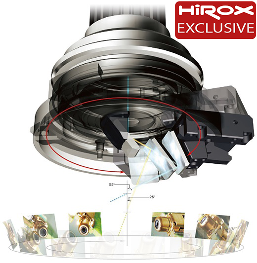
360 degree HIROX rotary optic head diagram
02

Rotary Head 360°

Automated lateral observation of edges, holes, welds, subframes and complex geometries.
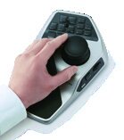
Remote control HIROX with joystick and touch screen
03

Complete remote control

Smart Joystick, touch screen and API to govern acquisition, movement and control recipes.
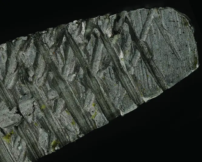
Comparison of multifocus 3D mapping and confocal profilemetry
04

Double 3D mapping

Optical Multi-Focus and NPS Nano Point Confocal Scanner to choose the most suitable method for the surface.
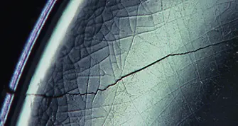
Step-by-step component entirely on fire during inspection
05

Infinite Auto‐Focus

Instant focus along the observed field, even during the exploration of the sample.
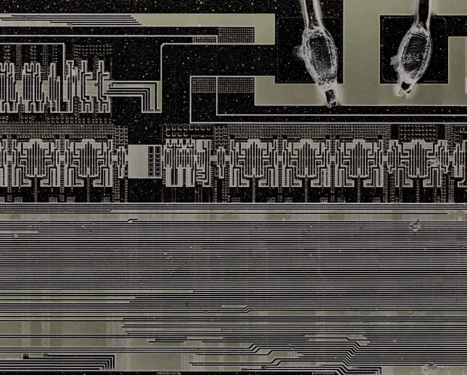
2D and 3D panoramic Stitching of a large electronic circuit
06

Big Picture / Infinite Stitching 2D/3D

High-resolution panoramic scans to move from the complete view of the sample to the single detail.
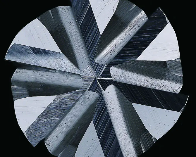
Methrological position with 3D analysis and automatic report
07

AutoReport + MountainsMap

Advanced metrological analysis and automatic generation of repeatable and shared reports.
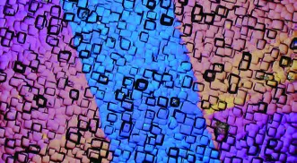
Multi-spectral comparison of the same sample in UV and NIR visible light
09

Multi-spectral acquisition

VIS, UV and NIR observation to highlight details and contrasts that cannot be read in the visible light alone.
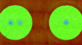
Selective angle lighting that highlights a superficial defect
10

Automated angle lighting

Controlled light management to bring out reliefs, engravings, defects and superficial morphologies.
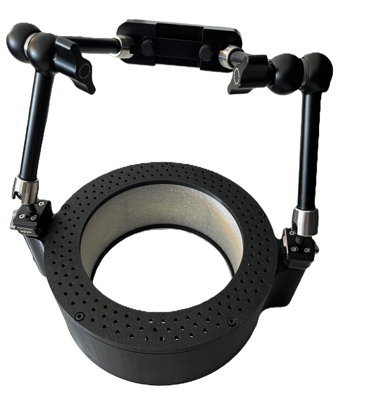
HIROX modular lighting system
11

Diffused lighting

J‐LED and J‐SFL systems to illuminate evenly complex, metallic and reflective samples.
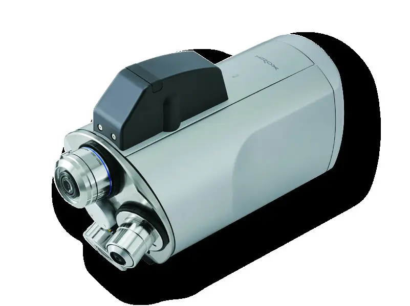
Z-Stack sequence transformed into image on fire and 3D reconstruction
12

AutoFocus and Z‐Stacking HR

High-resolution focal series combined in fully focus images and 3D reconstructions.
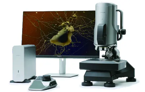
Original HIROX HRX-02 system
13

HiroxBot, calculation and CAD/FEM

Robotized inspection, contamination, reverse engineering, computational analysis and related 3D modeling.

PRODUCTS AND LIGHTS

Observe complex surfaces from normally inaccessible angles

360 degree HIROX rotary optic head diagram
HIROX EXCLUSIVE

360° motorized rotary optics

It allows you to observe subframes, holes, edges, grooves and lateral surfaces without constantly manipulating the component.

RVOMAX optical body

Integrate high-resolution chamber, macro lens 10–80×, mid‐range 100–500× and additional lenses to reach up to 10,000×.

Motorized telecentric zoom

Supports accurate 3D reconstructions even at low magnification, reducing prospect distortion.
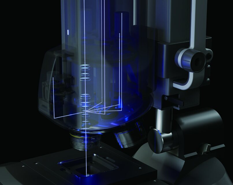
Hirox lighting

Selectable illumination

Light and dark field, coaxial, segmented, directional, diffuse, polarized, UV and IR for different materials and morphologies.

Configurable states

Manual or motorized axes, high precision Z axis and XY stages from 50 × 50 mm up to 1,500 × 1,500 mm.

HIROX HRX‐02 · OFFICIAL VIDEO

From 10× to 10,000× with the same platform

A visual overview of the HRX‐02 system, from optical configuration to observation and sample acquisition.

Open the HIROX video in a new window →
MULTI-FOCUS + CONFOCAL NPS

Two 3D modes, one analysis platform

The Multi‐Focus optical reconstruction quickly documents form and morphology; the NPS confocal sensor adds the contactless XYZ metrological measurement. The operator chooses the most suitable method for the surface and the required result.

HIROX HRX-02 system with NPS and three-dimensional surface display
HIROX MULTI-FOCUS

3D optical microscopy

The Multi‐Focus HIROX automatically captures a sequence of images at different odds along the Z axis.

The Z scan is carried out through the motorized and controlled movement of the stand. It does not require contact with the sample or its destructive preparation.

  • Completely on fire also on specimens with strong dislivelli.
  • Three-dimensional reconstruction of the surface.
  • 3D display in real colors, fake colors or wireframe.
  • Measures of height, profile, volume and roughness.
  • Analysis of defects, edges, engravings, cliques and complex geometries.
  • Digital documentation and repeatable reports.
01 · OBSERVATION

Multi-Focus optic

  • Combines a sequence of focal planes in a fully focusing image.
  • It quickly reconstructs morphology and visible altitude differences.
  • It is suitable for documentation, visual comparison and analysis of complex geometries.
02 · METROLOGY

White light confocal NPS

  • It acquires the contactless real height via confocal chromatic sensor.
  • Generate 3D profiles and surfaces with XYZ information.
  • Measure heights, distances, volume, roughness, ripple and topography.

Analytical surfaces

Specular, glossy, reflective, rough, opaque or transparent, selecting sensor and configuration according to the sample.

Combined result

High quality image to recognize detail and quantitative data to verify quotas, shape, defects and surface finish.

HIROX image example extended completely to focus
AUTO-FOCUS INFINITES · LIVE MULTI FOCUS

Explore the sample while keeping all levels readable

The HIROX system combines motorized control and real-time processing to overcome the limited field depth of the traditional microscope and clearly show surfaces with strong altitude differences.

01 · OBSERVATION

Focusing during the exploration

The operator can move to the sample and move from the general view to the detail, quickly recalling the sharp plane.

02 · EXTESA PROFONDITY

Live Multi Focus

When the sample has reliefs or differences, the HIROX standard function instantly generates an image completely on fire, without being bound to the depth of the field of a single frame.

03 · RESULTS

Details on different odds

  • Edges, steps, cliques and sloping surfaces easier to interpret.
  • Overall clear vision of three-dimensional components.
  • More immediate visual comparison between areas placed at different heights.
04 · OPERATIONS

Simple and repeatable acquisition

  • Controlled by the 4K HIROX interface.
  • Customizable and memorized settings for the sample.
  • Integration with acquisition, 2D/3D measurements and digital documentation.
1Pane the area of interest
2Define the quota range
3Get your vision fully on fire
3D digital microscope HIROX HRX-02

When it is particularly useful

Electronic components, processed surfaces, fractures, welds, finds, fibers and any sample in which more important details are found on different focal planes.

Official study HIROX HRX‐02 →
HIROX focal plane sequence combined in a three-dimensional reconstruction
HIROX · HIGH‐RESOLUTION Z‐STACKING

AutoFocus and Z‐Stacking HR

The system acquires a series of high-resolution images at different altitudes along the Z axis. The software selects the sharp zones of each floor and combines them in a single fully-focused image, which can also be used to reconstruct the 3D morphology of the surface.

1Definition of the Z range
2Automatic acquisition of focal planes
3Image fusion and 3D reconstruction
01 · ACQUISITION

High-resolution focal series

The motorized stand moves the lens along the Z axis with controlled increments, recording a consistent sequence of contactless sample images.

02 · FOCUS COMPOSITING

An image completely on fire

The result exceeds the depth of the field of the single floor, maintaining legible reliefs, cavities, edges and sloping surfaces.

03 · 3D MODEL

From the focal height to morphology

  • Three-dimensional reconstruction of the surface.
  • Real color display, fake colors or wireframe.
  • Virtual profiles and reading of height differences.
04 · ANALYSIS

Measures and documentation

  • Height, profile, volume, angle and radius.
  • Analysis of cracks, engravings, welds and complex geometries.
  • Repeatable acquisitions for comparison and reporting.
Three-dimensional display of a surface acquired with HIROX

Difference compared to Live Multi Focus

The Live Multi Focus facilitates the immediate observation of the sample. The Z‐Stacking HR instead favours a series acquired in a controlled manner, intended for high-resolution final images, 3D reconstructions, measurements and technical documentation.

HIROX HRX‐02: Live Multi Focus and 3D measures →
HIROX panoramic acquisition of a large electronic card
BIG PICTURE · INFINITE STITCHING 2D/3D · TILING & MAP

The complete sample becomes a navigable and measurable map

HIROX automatically combines adjoining acquisitions in a single high-resolution panoramic image. The general vision keeps the connection with every microscopic detail and allows you to quickly reach the area of interest.

01

Select the area

The operator defines the surface to be documented on the sample.

02

Automatic acquisition

The motorized XYZ displaces the sample and acquires the contiguous fields.

03

Infinite Stitching 2D/3D

The software aligns the fields and builds in real time a continuous overview, even with quota information.

04

Navigation on the map

Click the Big Picture to return the stand to the selected position.

From the overview to the microdetail

  • Wide field of view without sacrificing microscopic resolution.
  • Immediate localization of defects and areas of interest.
  • Comparison between distant areas of the same component.
  • Continuous documentation of larger samples of the single optical field.

Stored information

  • High resolution 2D panoramic image.
  • 3D map with geometry and altitude differences, when provided by the configuration.
  • Coordinates of the observed areas to find the same point.
  • Data available for measurements, annotations and reports.
Big Picture 2D of an electronic card
Big Picture 2DHigh-resolution panoramic mosaic to document, annotate, navigate and measure large surfaces.
Infinite 3D Stitching of an electronic circuit
Infinite 3D StitchingThe union of XY fields and Z acquisitions to correlate the extension of the sample to its morphology.
Electronic detail included in the Big Picture documentation
From the general picture to detailThe panoramic map keeps the spatial reference and allows you to quickly reach electronic details and areas of interest.
HIROX portal system for automatic acquisitions on large areas

Applications

PCBs and wafers, panels, mechanical components, coated surfaces, fabrics, documents, paintings and extensive samples requiring a detailed overview.

Official study HIROX: 3D Tiling & Map →
HIROX J-LED diffuser in ring shape
HIROX · DIFFUSION LIGHTING

Diffused lighting

A controlled light distribution around the sample reduces reflections and disuniformity, improving the quality of images and high resolution measurements.

J‐LED

Light annular 360 °

The new J‐LED diffusion lighting device works as a donut-shaped light box.

It is equipped with powerful LED lights that allow intense lighting on the whole sample to obtain high quality images.

Metal samples and other reflective materials can be difficult to acquire because they reproduce the light source on the surface. J‐LED distributes the 360° light around the sample, ensuring uniform illumination. It is particularly useful for complex samples such as metal ball bearings.

HIROX J-LED, diffused ring lighter
J-SFL

Flexible six-arm lighting

The new J‐SFL diffusion lighting device works as an openable light box with six arms.

It is equipped with 432 powerful LED lights, distributed on 13 controllable segments independently.The light direction can thus be adapted to the geometry and reflectivity of the sample.

HIROX J-SFL with six arms and segmented lighting
DOME LIGHTING

Dome lighting

For metals and reflective surfaces, the 360° light around the sample reduces the reflections of the source and distributes the illumination evenly.The solution is also suitable for curved geometries and samples difficult to observe with direct light.

Official page HIROX Europe: lighting →
HIROX rotary head operating principle with viewing angles from 25 to 55 degrees
HIROX EXCLUSIVE · 360 ° EXPENDITURE

Rotary Head 360°

The patented HIROX rotary head looks at the entire profile of the component without turning or repositioning the sample.

Motorized control

  • Rotation continues around the sample.
  • Speed, direction, zoom and angle selectable from the software.
  • Magnetic adapter automatically recognized by the HIROX system.

Observable geometry

  • Edges, holes, grooves and side walls.
  • Welding, subsquadri and details normally hidden.
  • Immediate comparison of the same detail from several directions.

Optical configuration

Observation angles
From 25° to 55° by means of a group of high precision mirrors.
Compatible lenses
HR‐2016E and HR‐5040E.
Indicative range
From 6× to 400×, depending on the optics and configuration.
Documentation
Fast acquisition of high-quality photos and videos.
HIROX DIMOSTRATIVE VIDEO

360° optical inspection

The rotating head looks at edges, side walls and subsquadri while keeping the sample steady.

Technical deepening HIROX Europe →
Ergonomic remote controller CT-R02 for HIROX HRX-02
CT‐R02 · ERGONOMIC CONTROL

Complete remote control

The remote controller concentrates the main HRX‐02 commands in an ergonomic position, simplifying observation and acquisition during repetitive tasks.

Controlable functions

  • Adjustment of illuminations.
  • System handling and sample navigation.
  • Motorized zoom control.
  • Quick capture of images.

Operating benefits

  • Main commands accessible without interrupting observation.
  • Faster adjustments during component inspection.
  • Increased continuity in acquisition and documentation sequences.
  • Intuitive interaction also in prolonged analysis.

Integration with HRX‐02

System
CT‑R02 remote controller for the HIROX HRX‑02 3D digital microscope.
Software interface
Physical commands support the 4K interface on Windows PC connected via USB.
Settings
The software allows you to configure and save custom settings for different samples and tasks.
Technical deepening HIROX Europe →

HIROX OTTLE GAMMA

From the macro-to-retail vision up to 10,000×

The HRX‐02 platform can be configured with motorized or manual optical groups. The choice depends on visual field, resolution, working distance, field depth and sample geometry.
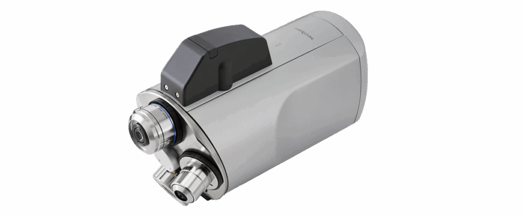
HIROX HRX-RVOMAX motorized optical body with door-objective tower
10×–10.000×

HRX-RVOMAX

Integrated unit with 5 MP room, motorized zoom, door-to-objective tower and built-in lighting.
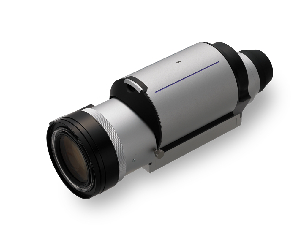
HIROX HR-1020E motorized telecentric lens
10×200×

HR‐1020E telecentric

High resolution motorized zoom, wide field of view and reduced perspective distortion: indicated for accurate 3D dimensional measurements and reconstructions.
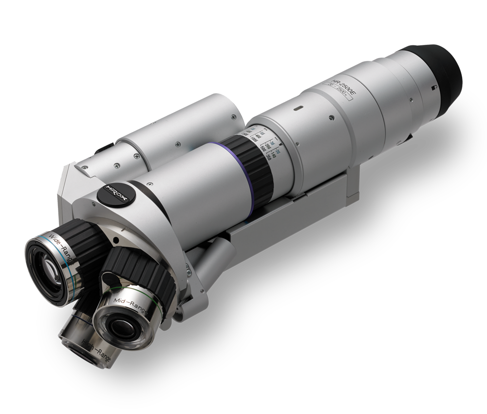
HIROX HR-2500E optics with three lens turret
20×5,000×

HR‐2500E / HR‐5000E

Revolver optics with three lens turret and double illumination, designed to quickly switch from the microscopy detail component.
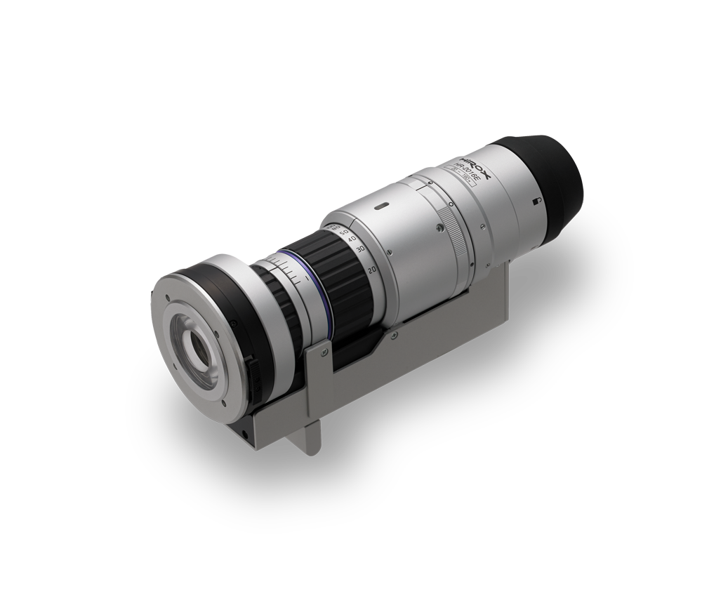
HIROX HR-2016E motorized optics with low magnification
6×320×

HR‐2016E · low magnification

Compact and versatile body for large samples, general control, documentation and applications requiring a wide field of view.
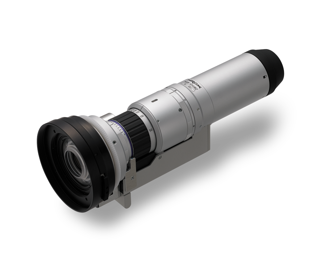
HIROX HR-5040E optics with medium magnification
20×–800×

HR‐5040E · medium magnification

Large depth of field and long working distance for irregular surfaces, three-dimensional components and operations that require space around the sample.
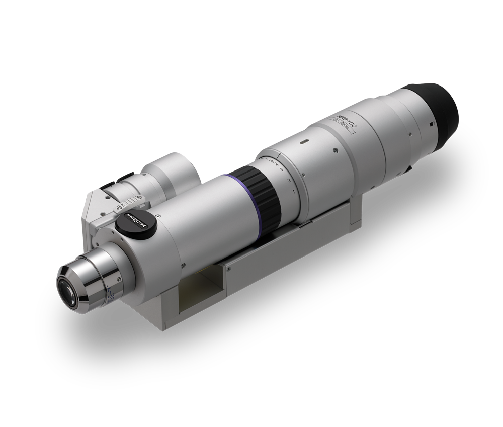
HIROX HR-10CE high resolution optics
35×–10.000×

HR‐10CE · High resolution

High magnification zoom with six interchangeable lenses for microstructures, particles, fine defects and analysis requiring maximum optical resolution.

HRX‐RVOMAX · OPTIC CONFIGURATION

From 10× to 10,000× with a single optical body

The motorized turret combines the Macro optics and the OLE‐100, OLE‐350, OLE‐500, OLE‐700 and OLE‐1000 lenses. The system switches from the general view of the sample to the analysis of the most fine details, maintaining the integrated management of zoom, focus and lighting.
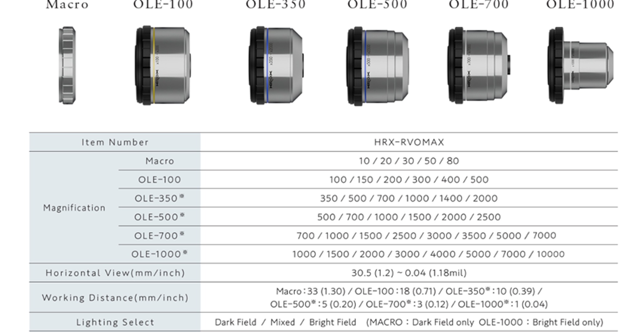
30,5–0,04 mm horizontal visual field33–1 mm working distance3 modes dark field, mixed and clear
HIROX HRX-RVOMAX system widening range and tableSelect the image to enlarge
Available magnifications and working distance
OpticsSelectable magnifiersWorking distance
Macro10× · 20× · 30× · 50× · 80×33 mm
OLE-100100× · 150× · 200× · 300× · 400× · 500×18 mm
OLE-350350× · 500× · 700× · 1,000× · 1.400× · 2.000×10 mm
OLE-500500× · 700× · 1,000× · 1,500× · 2.000× · 2.500×5 mm
OLE-700700× · 1.000× · 1,500× · 2.500× · 3.000× · 3,500× · 5.000× · 7.000×3 mm
OLE-10001,000× · 1,500× · 2.000× · 3.000× · 4.000× · 5.000× · 7.000× · 10.000×1 mm

Selectable lighting: dark field, combined and light field. Macro optics work in dark field; OLE‐100 in light field.

Manual versions

Available from 0.1× up to 10,000× for essential configurations or dedicated applications; the HR‐1020E is exclusively motorized.

Special optics

Borescopi, C‐mount attacks, BGA optics and customized solutions expand access to cavities, sub-squadri and out-of-standard applications.

Integrated lighting

The combination of annular, coaxial, directional, diffused and polarized light is selected according to reflectivity, texture and surface morphology.

HIROX HRX-RVOMAX
10×–10.000× · HRX‐02

HRX-RVOMAX

Compact unit with 2/3” high resolution room, fully motorized zoom and three-purpose turret. The lighting is integrated and controllable by the software without additional external cabling.

Optical and lighting

  • Macro optics, OLE‐100, OLE‐350, OLE‐500, OLE‐700 and OLE‐1000.
  • Overall horizontal field of view: 30,5–0,04 mm.
  • Working distance: from 33 mm to 1 mm, according to the objective.
  • Coaxial lighting / light field, dark and combined field.
  • Polarization, oblique light, fixed or central diaphragm and directional segmented ring.

Control and applications

  • Zoom, focus, turret and light managed by HIROX software.
  • Quick transition from general picture to high resolution.
  • Directional lighting to highlight reliefs, textures and superficial defects.
  • Configuration indicated for quality control, failure analysis, electronics, mechanics and materials.
Official HIROX Europe →
HIROX HR-1020E
10×–200× · TELECENTRICA

HR‐1020E

High resolution motorized telecentric zoom, designed to reduce prospect distortion and provide reliable dimensional results on a wide field of view.

Key data

Enlargement
10× · 20× · 30× · 50× · 90× · 100× · 120× · 150× · 200× ·
Working distance
54 mm
Horizontal visual field
30,5–1.52 mm
Motorization
Zoom

Magnetic adapters

  • AC‐1020C: coaxial lighting.
  • AC‐1020D: diffused lighting.
  • AC‐1020S: side light and contact inspection.
  • AC‐1020P: polarizer with filter slider.
  • Only motorized version.
Official HIROX Europe →
HIROX HR-2500E and HR-5000E
20×5,000× · TORRETTA TRIPLA

HR‐2500E / HR‐5000E

Revolver system with three optical ranges, double coaxial and annular lighting and motorized control of zoom, turret and light.

HR‐2500E

Gamme
20–140× · 140–1.000× · 350–2.500×
Working distances
18 · 10 · 10 mm
Visual field
15,4–2,21 · 2,18–0,31 · 0,87–0,12 mm
Field depth
0,72–0,072 · 0.09–0,007 · 0.04–0,002 mm

HR‐5000E

Gamme
20–140× · 140–1,000× · 700–5,000×
Working distances
18 · 10 · 3,4 mm
Visual field
15,4–2,21 · 2,18–0,31 · 0,43–0,06 mm
Lighting
Coaxial, annular and combined

Accessories

Directional, fixed, variable and central Diaphragms; coaxial polarizer; single wavelength filter; polarized dark-field ring; diffuser; lateral light and rotating adaptor.

Official HIROX Europe →
HIROX HR-2016E
6×–320× · BASSO INGRANDIMENT

HR‐2016E

Compact high depth field optics, which can also be used by hand and compatible with 360° rotating head and automatically recognized magnetic adapters.

Configurations

Standard
20–160× · WD 44 mm · FOV 15,4–2,0 mm
Low magnification
6–48× · WD 135 mm · FOV 45.5–5,84 mm
High magnification
40–320× · WD 20 mm · FOV 7,62–0,95 mm
Motorization
Zoom and rotary head

Main Adaptors

  • AC‐2016R and SP‐AC‐2016LOWR: 360° rotary inspection at 45°, without moving the sample.
  • AC‐2016S: contact and light at a variable angle.
  • AC‐2016H/HI: non-contact and high magnification.
  • AC‐2016D/VD: fixed or variable diffused lighting.
  • Magnetic connections with automatic recognition.
Official HIROX Europe →
HIROX HR-5040E
20×–800× · MEDIO INGRANDIMENT

HR‐5040E

Universal zoom with wide working distance and numerous fast graft adapters. The rotating head allows complete observations to multiple angles.

Configurations

Standard
50–400× · WD 54 mm · FOV 6,1–0,78 mm
Low magnification
20–160× · WD 80 mm · FOV 15,4–2,0 mm
High magnification
100–800× · WD 20 mm · FOV 3,05–0,39 mm
Motorization
Zoom and rotary head

Main Adaptors

  • AC‐5040RV: 360° rotating head, 25–55° angle, software control.
  • AC‐5040H/S: non-contact or contact.
  • AC‐5040D/VD: fixed or variable diffused light.
  • AC‐5040LOW/HI: 20–160× or 100–800×.
  • AC‐5040P: polarized lighting.
Official HIROX Europe →
HIROX HR-10CE
35×–10.000× · ALTA RESOLUTION

HR‐10CE

10× coaxial optical zoom with interchangeable lenses, developed for maximum resolution of the HR series and for an extensive range of high-enlargement observations.

Objectives and performance

OL-35
35–350× · WD 34 mm · FOV 9,83–1,05 mm
OL-70II
70–700× · WD 21 mm · FOV 4,42–0,47 mm
OL‐140 / 140II
140–1.400× · WD 30,5/12 mm
OL-350II
350–3.500× · WD 10,6 mm · FOV 880–90 μm
OL-700II
700–7.000× · WD 3,4 mm · FOV 400–40 μm
OL-1000
1,000–10.000× · WD 1 mm · FOV 300–30 μm

Lighting and adaptors

  • Coaxial vertical lighting and ACS function on all optics.
  • Directional light, fixed, variable and central diaphragms.
  • Coaxial polarizer and single wavelength filter.
  • Side light, rotating adaptor and dark-field ring.
  • Motorized zoom; motorized and manual versions.
Official HIROX Europe →

HIROX CONFIGUREMENT

Custom logs for each geometry and control process

From compact position to bridge and robotic systems: each tripod is designed and customised on the user's application needs.

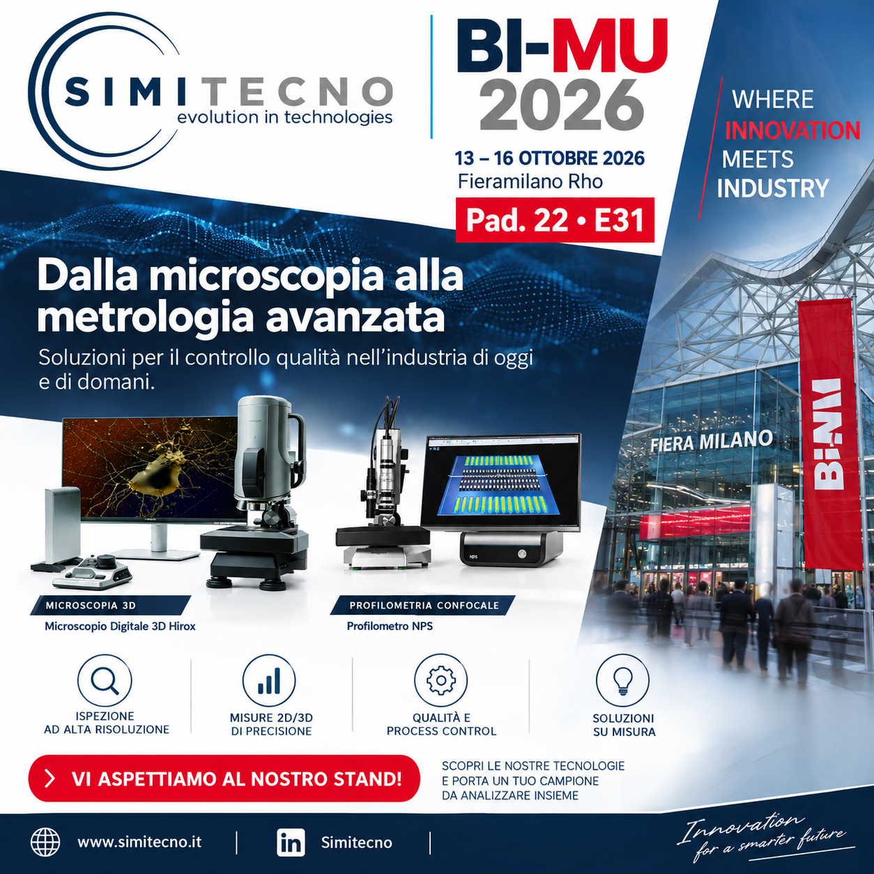
Complete Locandina Simitecno BI-MU 2026

PROJECT ON MEASURE

Mechanics, handling and electronics configured around the process

The number of motorized axes, the useful stroke, resolution, precision and speed of movement are defined according to the sample, the area to be inspected, the cycle time and the required automation level.

  • Motorized axesConfigurable number and cinema
  • RaceSized on the control area
  • ResolutionSelected according to the size
  • Speed speedOptimized compared to cycle time
  • Smart FactoryInterfaceable control electronics with PLC, MES and factory systems
HIROX

Stand range available

Standard and special configurations for HRX‐02 digital microscope and, where expected, confocal profile NPS.

ST‐AS‐XTilting counter stand up to 135°, fully motorized XYZ.
ST‐S‐XHigh precision vertical counter stand with motorized XYZ handling.
ST-ASIntelligent tilting stave with sensors, transmitted lighting and four controllable axes.
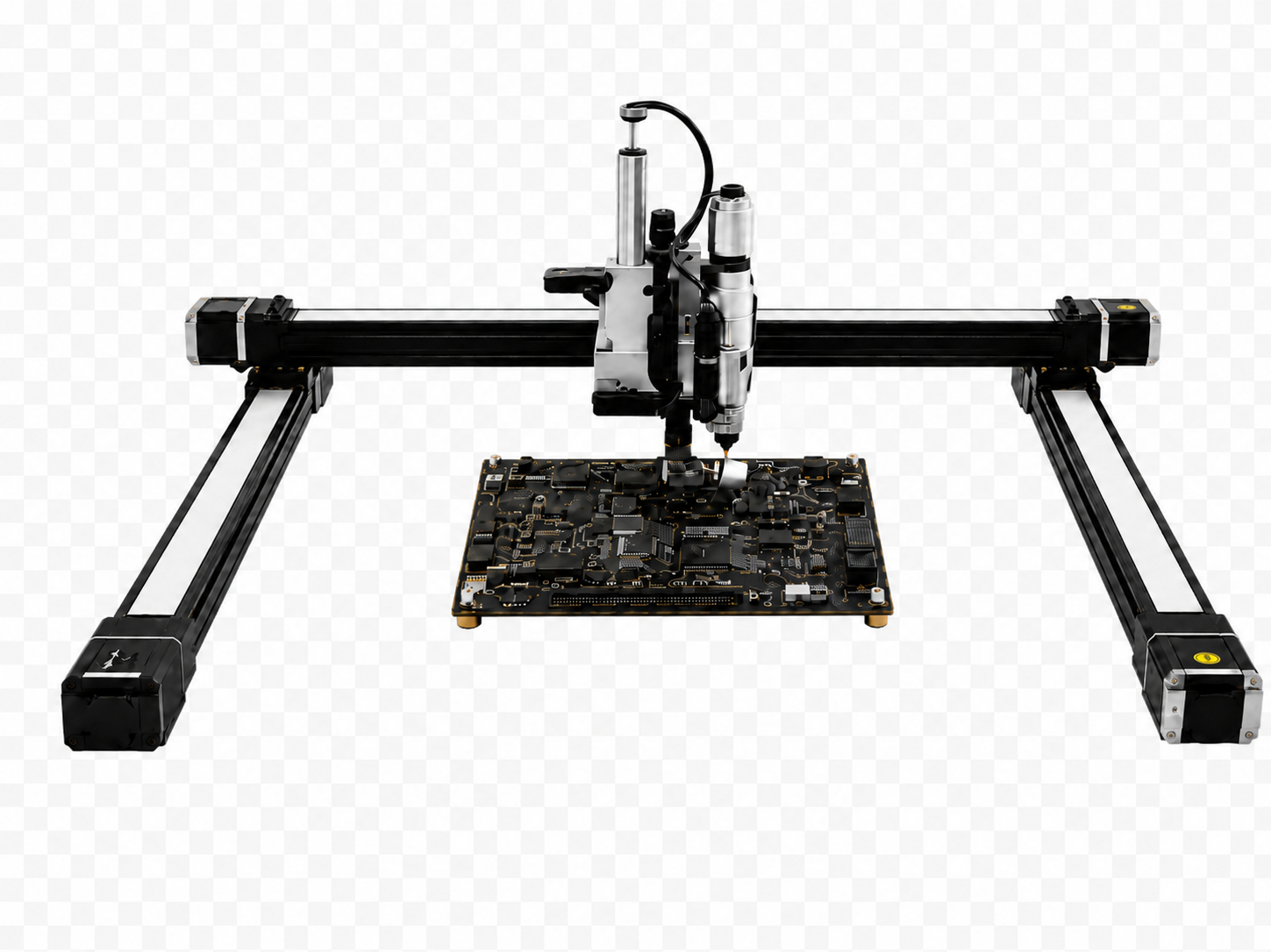
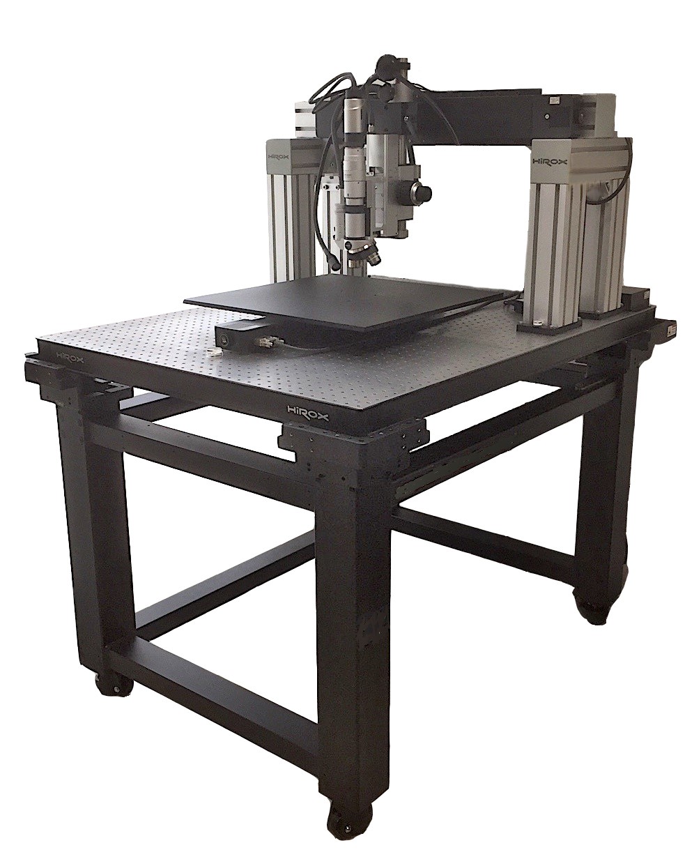
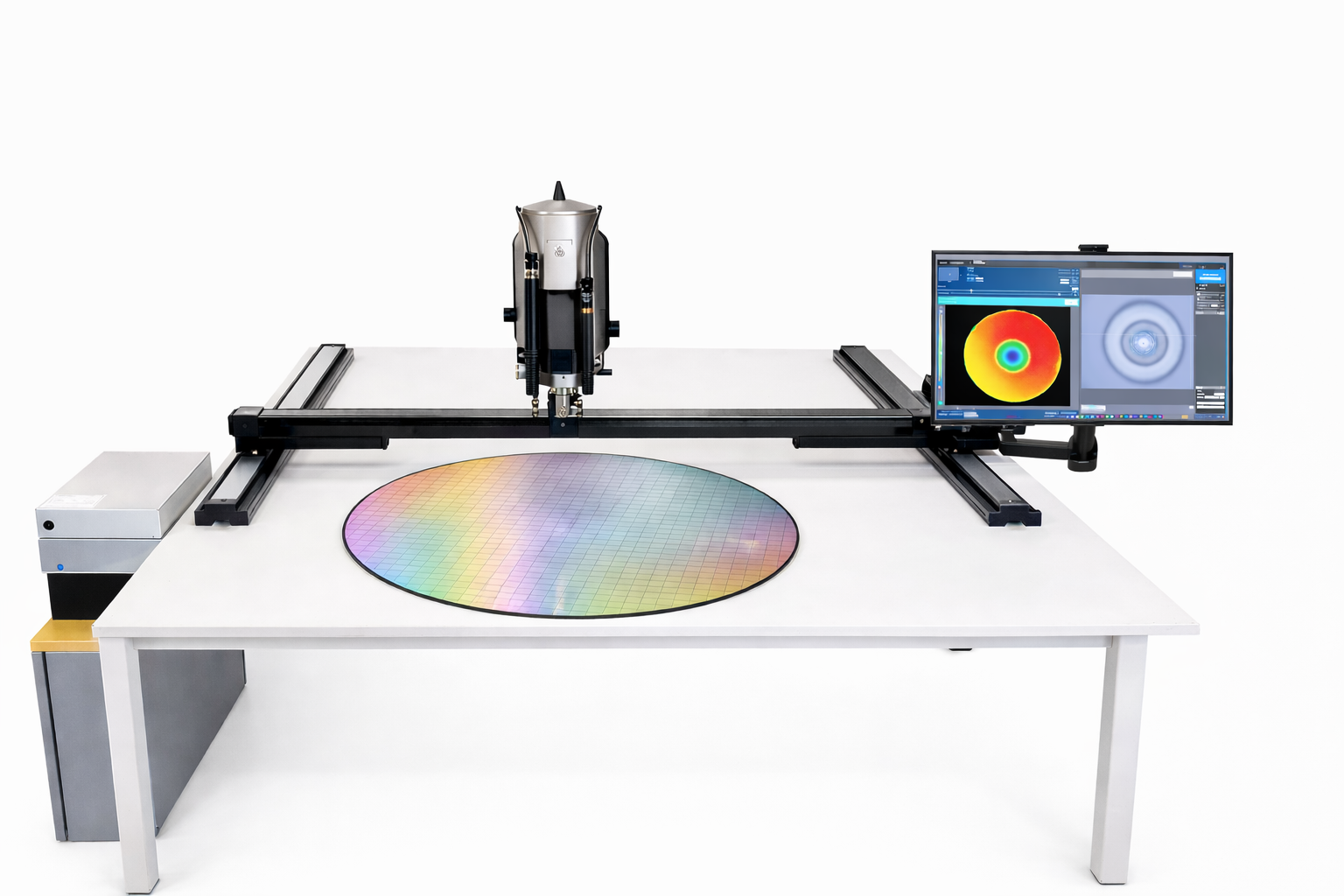
Gantry StandAutomatic surface scanning portal up to 1,500 × 1,500 mm.
BridgeMotorized bridge for plates, PCBs and samples extended up to 800 × 800 mm.
T‐StandardXYZ horizontal structure for walls, paintings, statues and other vertical objects.
Flexible Arm StandAdjustable column and flexible arm to observe 3D objects from any side.
ContactPortable contact stand for plates, cylinders, pipes and safety print.
Special StandsTailored systems for specific geometries, races and automations.
01

XY portals and systems for large areas

Extended rigid structures and racing for mapping, stitching and automatic control of panels, PCBs, voluminous works and components.
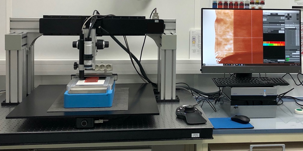
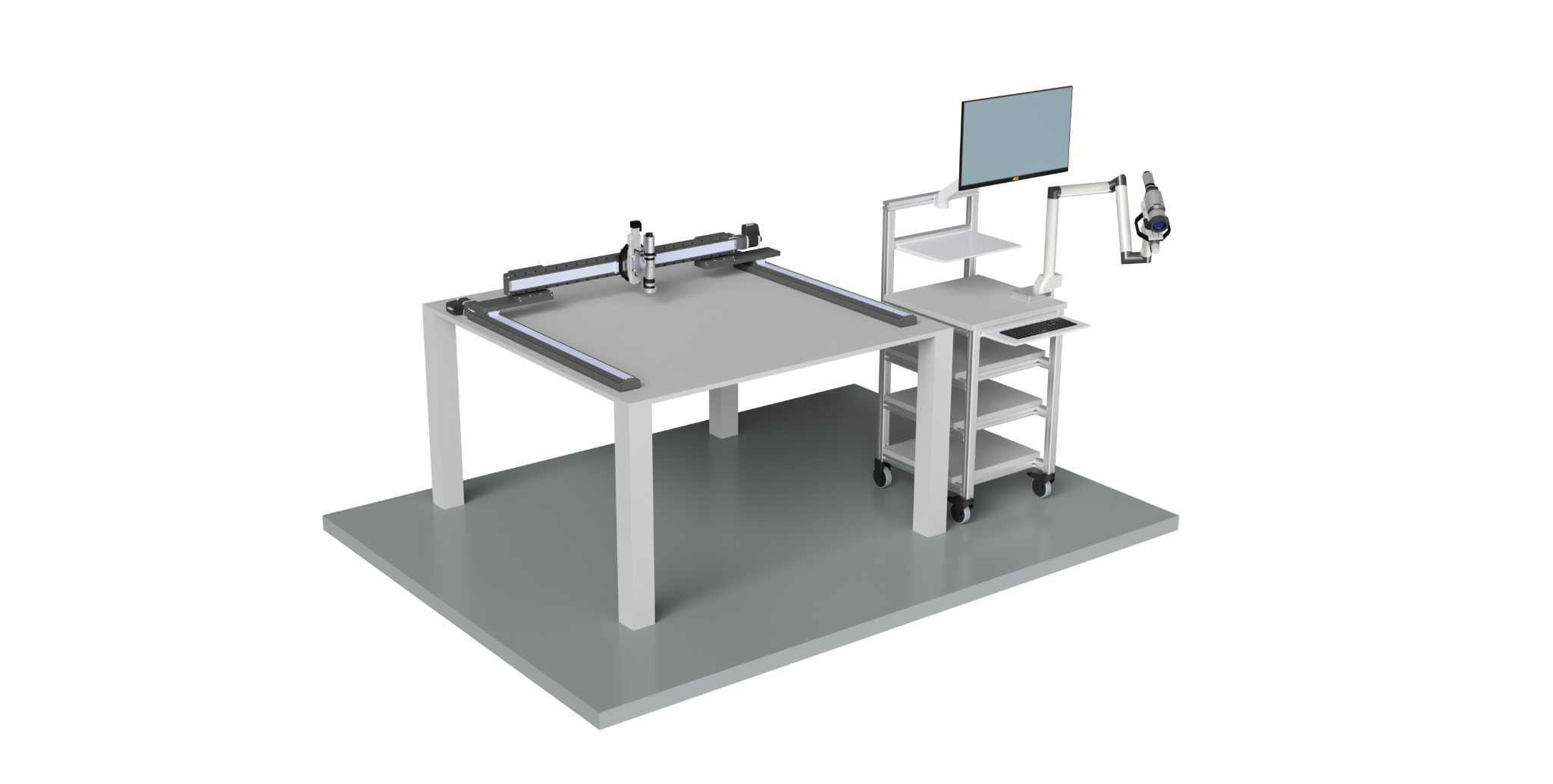
HIROX portal with XY table
XY Lab PortalPlanned scanning of flat surfaces with real-time analysis and measurement.
Large Size HIROX System
Customized measuring islandCustom Layout for extended samples and integration of multiple sensors.
HIROX Simitecno portal
Automatic control doorMotorized axes and repeatable recipes for serial controls on defined areas.
Large HIROX Bridge Stand
High stability bridgeReinforced structure for repeatable measurements on heavy or bulky samples.
HIROX portal for large surface
Gantry for large surfacesExtended XY stroke for high resolution mosaics and inspection of large areas.
HIROX optical system on linear guide
Dedicated linear axisTargeted handling for profiles, long samples and automatic sequences.
02

Collaborative robots and automatic cells

Flexible configurations to reach multiple sides of the piece, store the control points and integrate the microscope into the production line.
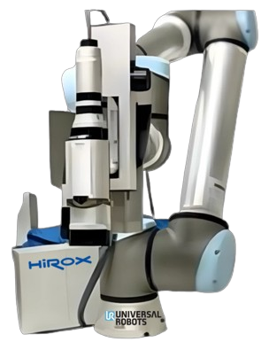
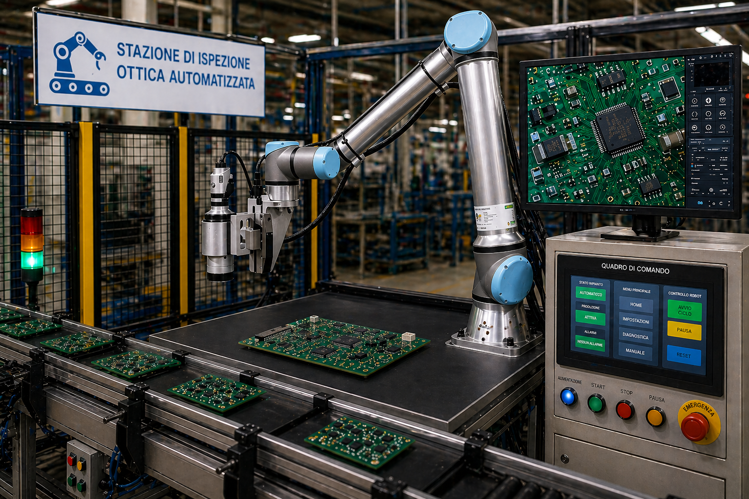
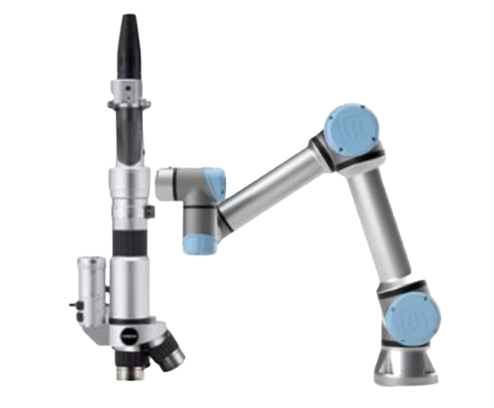
HIROX Microscope on Cobot
Cobot with HIROX opticalFlexible positioning of the optics around complex components.
Inspection Robotic Cell
Cella Smart FactoryAutomatic inspection with HMI, safety and connection to factory systems.
Manual and robotic HIROX configurations
Modular ConfigurationThe same platform can operate manually, on stand or on cobots.
Mobile Inspection Cobot
Cobot on a mobile basisTransferable station between different lines and control areas.
HIROX optics on robotic arm
Articulated robotic armAutomatic access to lateral surfaces, inclined and difficult points.
HIROX system on cobot with counter
Complete Robotic BankCobot, sample and software gathered in an integrated station.
03

Compact, manual and special statistics

Bench or transportable solutions for quick controls, local samples and measures that require great accessibility.
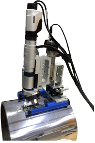
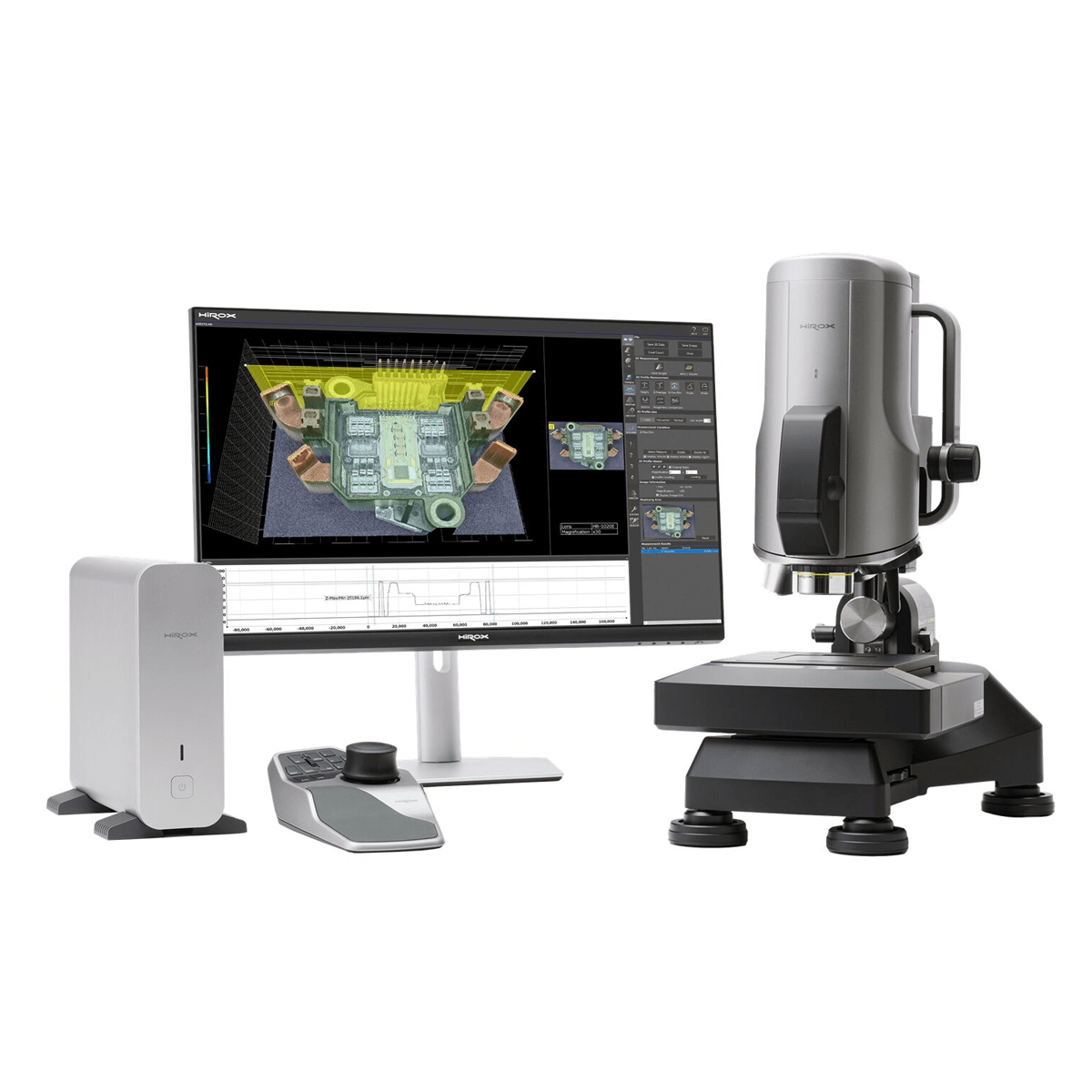
HIROX cable for cylindrical surface
Dedicated Compact StaticTargeted configuration for curved surfaces and hard-to-place parts.
HIROX Microscope on Bench Stand
Vertical counter standCompact solution for multifocus and 2D/3D measurements daily.
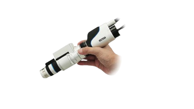
HIROX optics with manual handle
Portable optics with handleOn-site inspections directly on the component, without stands or supports.
Industrial HIROX special
Special industrial statisticsConfigurable robust structure for heavy parts and out-of-standard applications.
Gantry HIROX for large surfaces
SUPERFICIAL GRANDS · XYZ MOTORIZED

Gantry

Portal system to acquire, measure and reconstruct in 2D/3D very extensive samples, keeping the area free and accessible.

Key specifications

Racing X/Y
Up to 1,500 × 1,500 mm
Race Z
75 mm
Step of movement
Less than 0.2 μm
Sizes available
100, 200, 300, 500, 800, 1,000 or 1,500 mm

Functions and uses

  • Motorized control of X, Y and Z axes.
  • Compatible with multifocus, 2D/3D measurements and gigapixel stitching.
  • Suitable for large-format paintings, fabrics, panels and flat objects.
  • Mobile station available as an option.
Technical data: HIROX Europe range
HIROX tripod
ALTA STABILITY · XYZ MOTORIZED

Bridge stand

Rigid bridge architecture for automatic inspection of plates, electronic boards and large plane components.

Key specifications

Racing X/Y
Up to 800 × 800 mm
Race Z
75 mm
Step of movement
Less than 0.2 μm
Sizes available
100, 200, 300, 500 or 800 mm

Functions and uses

  • Motorized movement X/Y/Z.
  • Multifocus, 2D/3D measurements and high resolution stitching.
  • Configuration suitable for PCBs, plates and electronic components.
  • Mobile station available as an option.
Technical data: HIROX Europe range
HIROX stand
ST‐AS‐X / ST‐S‐X / ST‐AS

Motorized banknotes

Three high-precision laboratory platforms: vertical, fully motorized or inclined intelligent with sensors.

ST‐AS‐X and ST‐S‐X

Racing X/Y
Up to 200 × 100 mm
Race Z
75 mm
Step of movement
Less than 0.2 μm
ST‐AS‐X Inclination
Up to 135°

ST-AS

  • Motorized XY table from 50 or 100 mm; optional manual table.
  • Motorized Z stroke of 80 mm and pitch less than 0.2 μm.
  • X/Y/Z control/rotation via wireless 3D mouse.
  • Transmitted lighting, antivibrating feet and sensors of height and angle.
Technical data: HIROX Europe range
Compact HIROX for curved surfaces
PORTATIA · CURVE SUPERFICI

Contact stand

Compact support to be placed directly on plates, cylinders and pipes when the sample cannot be brought under a traditional static.

Key specifications

Race Z
30 mm
Step Z
0.05 μm
Table XY
Manual Micrometrics included
Motorized XY
Optional, 50 or 100+ mm

Applications

  • Inspection on the place of non-moving components.
  • Suitable for flat, cylindrical and tubular surfaces.
  • Allows fine focus and controlled positioning.
  • Configurable according to geometry and area to be examined.
Technical data: HIROX Europe range
HIROX optics with manual handle
PORTATIO · INSPECTIONS ON SITE

Optics with manual handle

Handheld configuration to bring the optics directly to the area to be controlled, without using stands, columns or other supports.

How to use

  • The operator manually supports and directs the optics through the handle.
  • Direct control of the component in the place where it is installed.
  • No support or static plan required.
  • Suitable for bulky, fixed or non-transportable samples.

Application advantages

  • Maximum freedom of positioning and orientation.
  • Access to vertical, inclined surfaces and areas difficult to reach.
  • Quick passage between different inspection points.
  • Suitable for production, maintenance and field inspections.
HIROX Portable Configuration
HIROX T-Stand for vertical surfaces
VERTICAL SUPERFICI · XYZ MOTORIZ

T‐Standard

Motorized horizontal stave to perform scans on paintings, walls, statues and other vertical objects without moving the sample.

Key specifications

Racing X/Y
Up to 1,500 × 1,500 mm
Race Z
75 mm
Step of movement
Less than 0.2 μm
Sizes available
100, 200, 300, 500, 800, 1,000 or 1,500 mm

Functions and uses

  • Motorized XYZ movement in front of the vertical surface.
  • 2D acquisitions, multifocus and 3D stitching over long distances.
  • Suitable for conservation, research, museums and large artefacts.
  • Mobile station available as an option.
Technical data: HIROX Europe range
HIROX stand with flexible arm
OGGETTI 3D · MASSIMA ACCESSBILITY

Flexible Arm Stand

Stable stave with adjustable column and flexible arm, designed to observe and measure flat, vertical, inclined or voluminous objects.

Configuration

Movement XY
Manual by handle
Race Z
75 mm, motorized
Installation
Fast assembly and disassembly

Applications

  • Inspection from different sides and perspectives.
  • 2D/3D measurements on findings, mechanical parts and bulky objects.
  • Also suitable for walls and surfaces difficult to reach.
  • Configuration distinct from the palm optics without support.
Technical data: HIROX Europe range
Special and automated HIROX system
PROJECT ON MEASURE · AUTOMATION

Special stands

Customized systems for samples, races and out-of-standard control flows, including portals, dedicated axes and robotic integrations.

Customisation

  • Dimensions, number of axes and kinematics defined on the application.
  • Support for HRX‐02 and NPS systems.
  • Acquisition, 2D/3D measurements, multifocus and configurable automation.
  • Custom software and integration into the customer system.

Possible configurations

  • Cells with collaborative robots and access to multiple sides of the piece.
  • Linear portals and guides for extended areas or profiles.
  • Mobile or integrated positions in the production line.
  • Performance and racing are defined in the project phase.
Technical data: HIROX Europe range

SOFTWARE 4K

From image to metrological data

Observation settings can be stored and recalled, increasing speed and repeatability. Encoded lenses and identified accessories reduce configuration errors.

Try these functions on the sample
01

Live HDR

Automatic optimization of brightness, contrast and exposure even on difficult surfaces.

02

Live Multi Focus

Fully focus images of objects with strong height differences.

03

Stitching 2D and 3D

Automatic union of adjacent areas to create high-resolution navigable maps.

04

2D Measures

Length, diameter, perimeter, angle and area with automatic calibration.

05

Metrology 3D

Profiles, heights, distances, volumes, roughness, rays and virtual sections.

06

Particle analysis

Automatic detection, classification and pass/fail results with statistical data.

APPLICATIONS

Configured around your component

DOESN'T YOU KNOW WHAT CONFIGURATION TO DECID?

Bring your sample to the Demo Center.

We will evaluate the most suitable optical, lighting, static and measuring method.

Book the HIROX test
← Back to products

CROMATIC CONFOCAL PROFILOMETRIES

NPS Nano Point Scanner

Submicrometric measurements of profiles, shapes and surfaces without contact, even on transparent, reflective or mirrored materials.

Request a NPS test
NPS Nano Point Scanner
76 nmAccuracy Z up to
2,000 points/sAcquisition of height
150–24,000 μmFull range of sensors
ISO 25178-602Color confocal profilemetry

METROLOGY AND MICROSCOPY

Two related tools on the same sample

The HIROX digital microscope quickly detects the area of interest and documents the high-resolution sample. The NPS sensor then performs the contactless confocal measurement on the same area, transforming the wavelength reflected into a Z share.
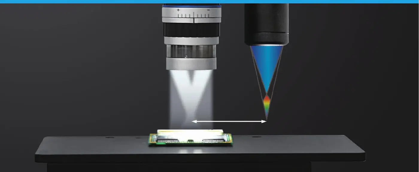
  • Opaque, glossy, reflective or transparent samples
  • Measurements in ambient light, without destructive preparation
  • Transition from optical vision to 3D metrology
HIROX digital microscope and NPS sensor on the same sample
Digital microscopy and integrated confocal profilemetry.

MAIN FEATURES

Non-contact 3D profilemetry, from roughness to shape

NPS combines a chromatic confocal point sensor, a high-precision XY stage and HIROX Maps metrological software. The configuration is chosen according to the Z field, the working distance, the slope of the surface and the required resolution.

Any surface area

Measure opaque, glossy, mirrored, rough or transparent materials without contact with the sample.

Profile and surface

Acquisition of single, programmable multi-profiles and high-resolution XYZ maps.

Large areas

Configurable motorized staves and boards up to 500 × 500 mm for flatness, undulating and composing.

Automatic control

Reusable templates, tolerance limits, pass/fail outcome and exportable results for traceability.
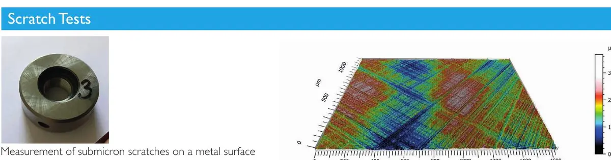
NPS three-dimensional profile of a superficial defect
From the visible defect to the quantitative measure of depth, shape and volume.
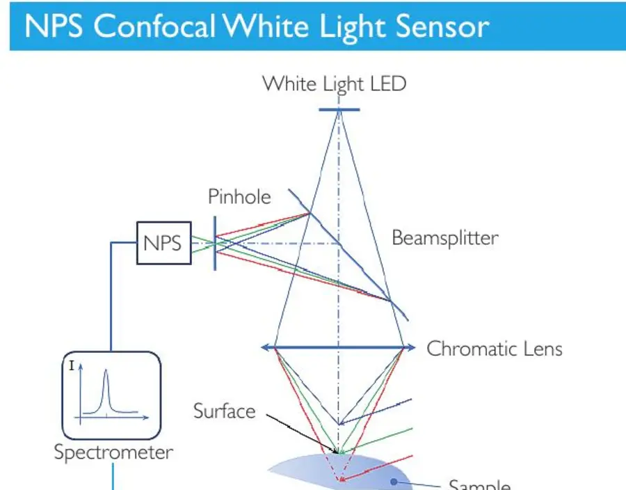
Colorful NPS Confocal Sensor Scheme
Optical diagram of the confocal sensor with white light.

PRINCIPLE OF MEASURE

From white light to XYZ

  1. Projection. The white light LED crosses the beamsplitter and the chromatic lens.
  2. Focalization. Each wavelength is focused on a different height on the surface.
  3. Confocal selection. The pinhole isolates the only perfectly focused wavelength.
  4. Conversion. The spectrometer translates the wavelength into height information.

The controlled movement of the XY stage turns the individual points into a linear profile or into a three-dimensional surface.

MODE OF ACQUISITION

Quick profile to 3D topography

The scanning strategy is chosen according to the geometry, area and parameter to be controlled.

01

High speed profile

Scan on a board for height, distance, radius, angle and linear roughness Ra, Rz and Rt.

02

Programmable Multiprofile

Multiple sections acquired in sequence and gathered in a single comparable report.

03

High resolution surface

Lined profiles generate XYZ data for Sa, Sz, volume, shape, wave and topography.

04

Form and large areas

Planarity, modularity, deformation and free geometries with internships up to 500 × 500 mm.

CONFIGURATION MODULATION

A sensor for each measurement scale

The most contained field favours accuracy; larger fields allow to measure higher components and extended forms.
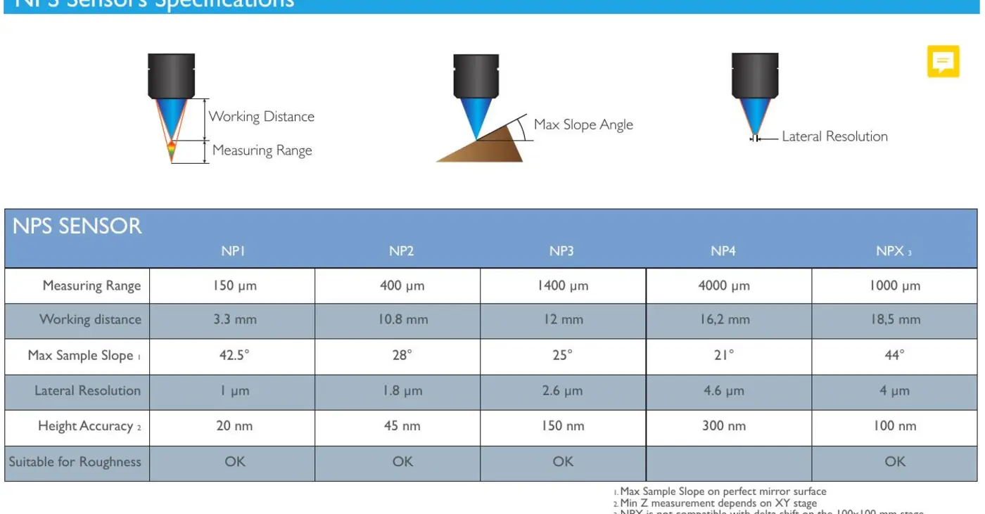
Comparative chart and table of NPS NP1 NP2 NP3 NP4 and NPX-1 sensors
Selection parameters: field, working distance, slope, side resolution and accuracy Z.
SensorCamp ZWorking distanceMaximum pendenceLateral resolutionAccuracy Z*Roughness
NP1150μm3,3 mm42°1.3 μm76 nmYes.
NP2400 μm10.8 mm28°1.7 μm169 nmYes.
NP31.400 μm12.2 mm25°2,6 μm500 nmYes.
NP44,000 μm16,5 mm21°7 μm1.195No.
NP512,000 μm26,6 mm14°11 μm3.350 nmNo.
NP624,000 μm20 mm8,5°11 μm6,850 nmNo.
NPX‐11,000 μm18,5 mm44°2.5 μm334 nmYes.
NPX‐22,000 μm19.2 mm42°3,8 μm675 nmYes.
NPX‐66,000 μm13,7 mm32°5.2 μm1.750 nmYes.

*The height accuracy also depends on the accuracy of the XY table. The maximum gradient is based on a perfectly speculative surface; additional sensors and configurations are available upon request.

NPS + HIROX MAPS

Automatic measurement, analysis and reports

The NPS software controls stage, sensor, frequency, light intensity and points of interest. The raw data is processed in Hirox Maps with Mountains technology for advanced analysis and repeatable procedures.

Live acquisition

Profile or map in false colors displayed during scanning.

Data correction

Leveling, removal of form, management of unmeasured points and extraction of areas.

Metrology Template

Reusable analysis sequences on samples and subsequent batches.

Pass/fail and export

Automatic tolerances, statistics and export CSV, Excel, STL and 3D formats.
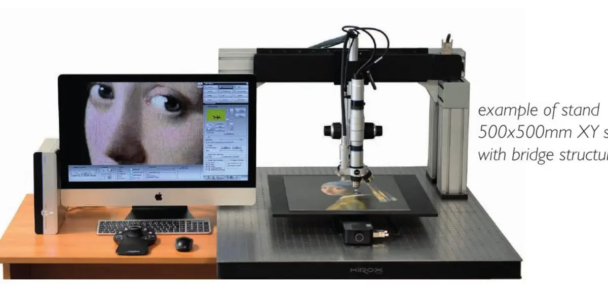
NPS system on a bridge with large XY internship
Bridge configuration for specimens and extended measuring areas.

REAL APPLICATIONS

3D measures for research, laboratory and process control

Each image shows the link between sample, contactless acquisition and metrological result.
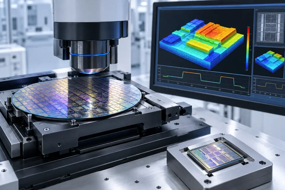
NPS measurement of wafers and microstructures for semiconductors
01

Semiconductors

Step height, thin films, wafer flatness and topography of microfabricated structures.
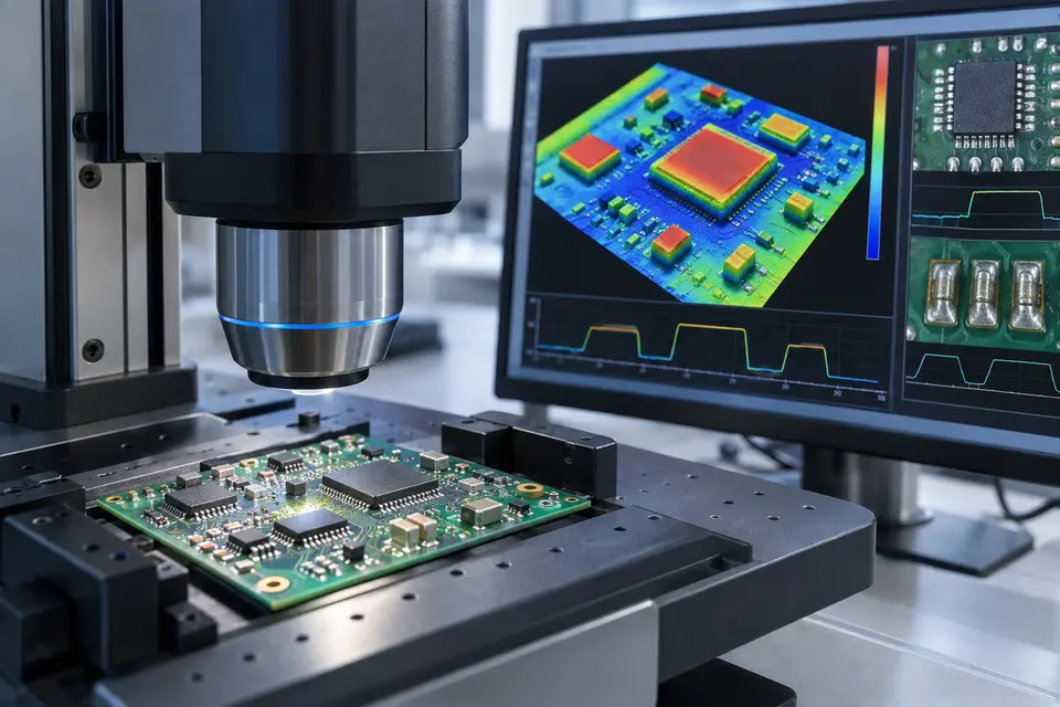
NPS control of a microelectronic board
02

Microelectronics

Height of components, textures, welds, pads, pistes and surface defects.
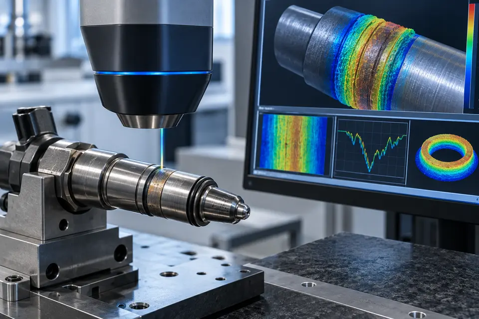
NPS profile of an automotive component
03

Automotive

Roughness, wear, sealing surfaces and geometry of precision machined components.
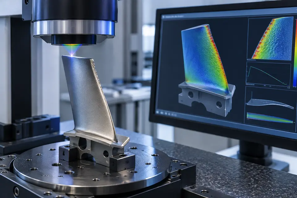
NPS measurement of aerospace shovel
04

Aerospace

Profiles, edges, coatings, curvature and damage of critical components.
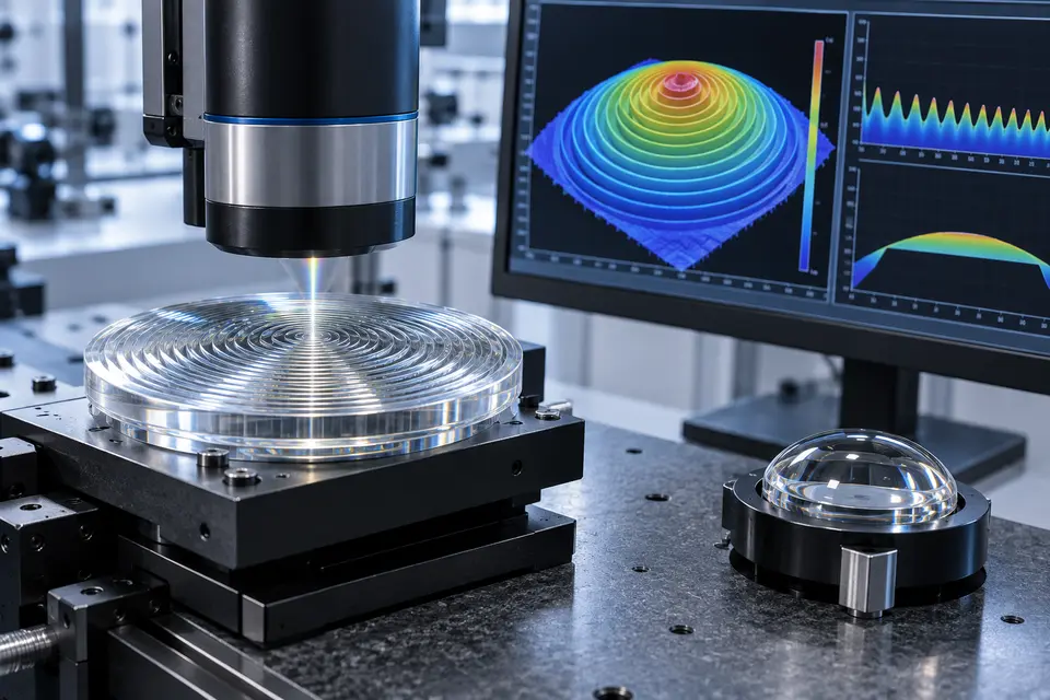
NPS measurement of Fresnel lens and transparent optical component
05

Optics

Fresnel lenses, transparent or mirrored surfaces, shape, radius and microstructures.
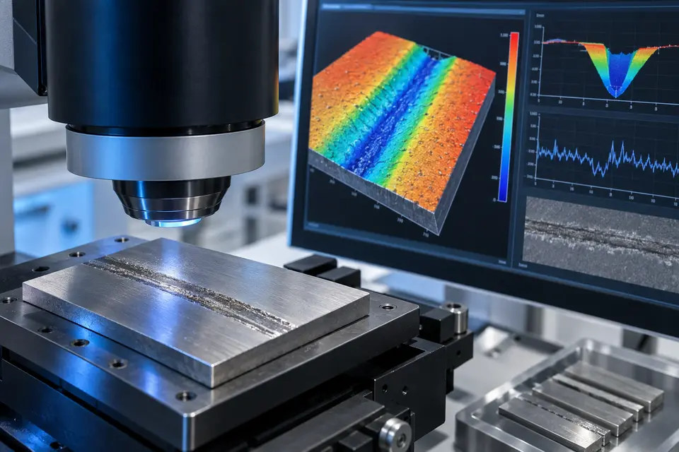
Tribological wear track NPS analysis
06

Tribology

Depth of the grooves, removed volume, roughness and characterization of wear tracks.
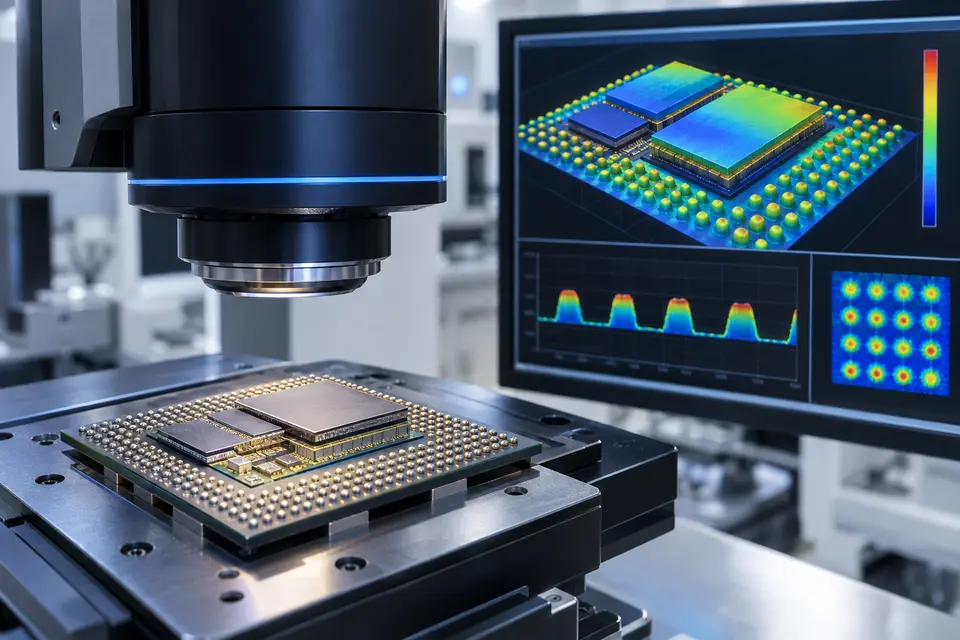
3D Microelectronic Packaging NPS Control
07

Packaging 3D

Micro-bump, interposer, stacked dies, height and complacency of interconnections.
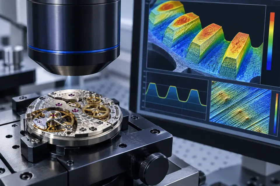
NPS profile of a watchmaking movement
08

Clockwork

Gears, teething, engravings, finishing and wear of movement components.

PROFILOMETRIES WITHOUT CONTACT

We verify resolution, field and sensor on your sample.

Book the NPS test

Technical data verified on Official HIROX Europe tab of Nano Point Scanner. The configuration and final performance must be confirmed according to sensor, table and application.

← Back to products

TARAZ METROLOGY · AREAL SURFACE

ASM — 3D areal surface measurement

Optical projection of fringes in blue light to quickly acquire shape, topography and roughness of metals, polymers and composites, without contact with the sample.
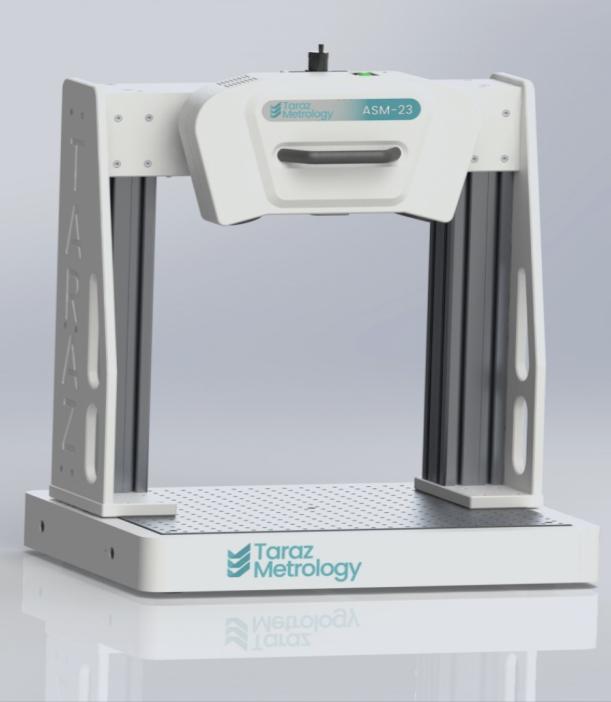
Taraz ASM system for 3D range measurement
4measurement configurations
1 μmLateral resolution
< 2Acquisition
ISO 25178analytical analysis

FROM LABORATORY TO LINE

A platform, four control ladders

ASM is the most advanced Taraz system based on blue light fringe projection. The non-contact optical measurement produces a full 3D point cloud of the surface and can be configured for off-line controls, automatic stations or online integration.

The same architecture covers precision components, processed surfaces, coatings and large panels, maintaining a common software flow with TarazSurf.
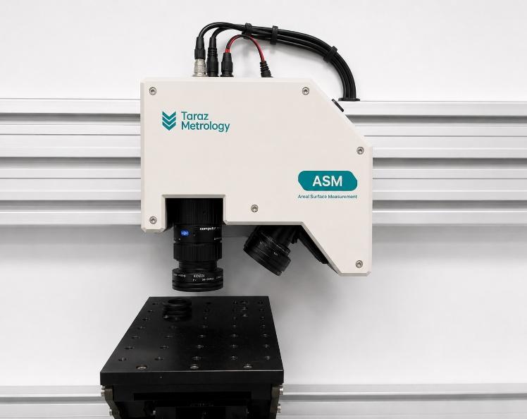
  • Quick and adjustable measurement of metals, polymers and composites
  • Complete traceability according to NPL metrological guidelines
  • Software and integration that can be adapted to the component and process
Taraz ASM Fine Resolution optic head on measuring board

GAMMA ASM 2026

The correct configuration for field and detail

All systems generate points clouds in .ply format and include TarazSurf software.

VersionCloud Point ResolutionMeasuring areaAcquisitionPoint cloud
Standard Resolution70 μm255 × 174 mm26
Higher resolution25 μm70 × 50 mm26
Wide-field400 μm1.200 × 800 mm26
Fine. NEW1 μm10 × 10 mm< 26
ENVIRONMENT22–30 °C

Declared operating range.

OUT.Point Cloud .ply

3D data ready for analysis and integration.

SOFTWARETarazSurf included

Acquisition, analysis and flow common to the range.

CONFIGURATIONLaptop available

Off-line, automatic and in-line solutions.

NEW · END RESOLUTION SYSTEM

Control of precision finishes

The new variant extends the ASM platform to a lateral resolution of 1 μm on a range of 10×10 mm. It is designed for the inspection of polished, lapped and finely machined components, maintaining a rapid, optical and safe measurement even on delicate surfaces.

1 μmLateral resolution10 × 10 mmArea field< 2Measuring time
ASM Fine Resolution System for polished and lapped surfaces

APPLICATIONS

From microfinish to large surfaces

The selection of the version depends on the area to be covered, the scale of detail and the cycle time required.

01

Automotive

Processed surfaces, precision components, wear, texture and process control.

02

Coatings

Topography, uniformity, finishing and defects on coatings and treated surfaces.

03

Additive manufacturing

Roughness, morphology and compliance of the surfaces produced by additive manufacturing.

04

Aerospace

Quick and traceable control of critical surfaces and components.

05

Precision finishings

Polished, lapped and finely machined components with Fine Resolution.

06

Large panels

Wide-field acquisition up to 1,200×800 mm in one measurement field.

Light Frange projected on a component during optical measurement

TARAZSURF

Measurement, analysis and traceability in a single flow

01

Optical acquisition

The blue light fringe projection detects the contactless surface.

02

3D Point Cloud

The system generates the complete data in .ply format.

03

Area analysis

TarazSurf supports surface analysis according to ISO 25178.

04

Integration

Results and procedures can be adapted to the part and process of the customer.
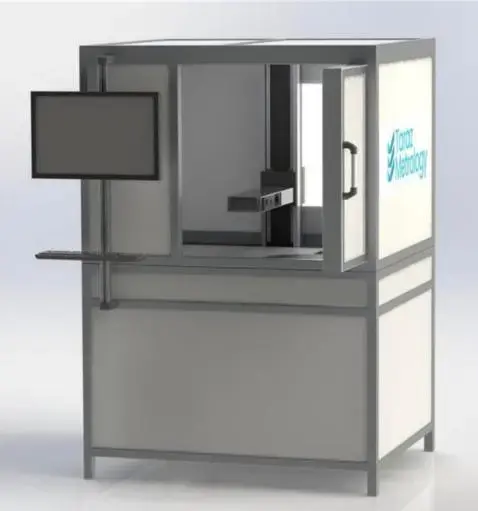
WHAT ASM FOR YOUR SUPERFICIE?

We verify field, resolution and cycle time directly on the sample.

Request an ASM test
← Back to products

TARAZ METROLOGY · POST-PROCESS MEASUREMENT

PM — 3D optical measurement after machining

Methrological station for final control of complex components in workshop and production. Integral frange projection, short-range photogrammetry and multiple viewpoints to achieve a full three-dimensional measure.
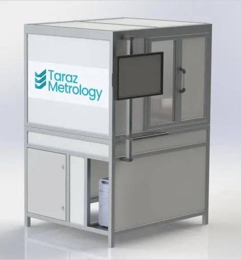
Metrological station Taraz PM closed
3D from multiple viewscomplete reconstruction
CADcomparison and deviations
Frange + photoscombined technologies
Productionfinal control

FINAL CONTROL

Measure the shape from multiple directions

PM acquires the component from different orientations and combines the measurements in a single 3D model. The precise tracking of the handling allows you to connect each view to the same reference system, while the recomposition algorithms merge the data of the different acquisitions.

The result can be compared with the nominal CAD model to immediately make the shocks visible from the tolerances and locate the areas that require verification.

  • Components with curved surfaces, subframes and articulated geometries
  • Adaptable configuration to size, shape and material of the piece
  • Independent station for post-process inspection
Taraz PM open with accessible measuring area

COME FUNZIONA

From the acquisition from several views to the scaling map

The cycle is configured on the component and the metrological target.

01

Positioning

The component is placed on the rotary table at the measuring station.

02

Optical acquisition

Screening of fringes and photogrammetry detect form and references from multiple viewpoints.

03

Data fusion

Tracing the movement and recomposition of data gather acquisitions in a single 3D measure.

04

CAD Comparison

Nominal deviations and tolerances are highlighted to support the decision.
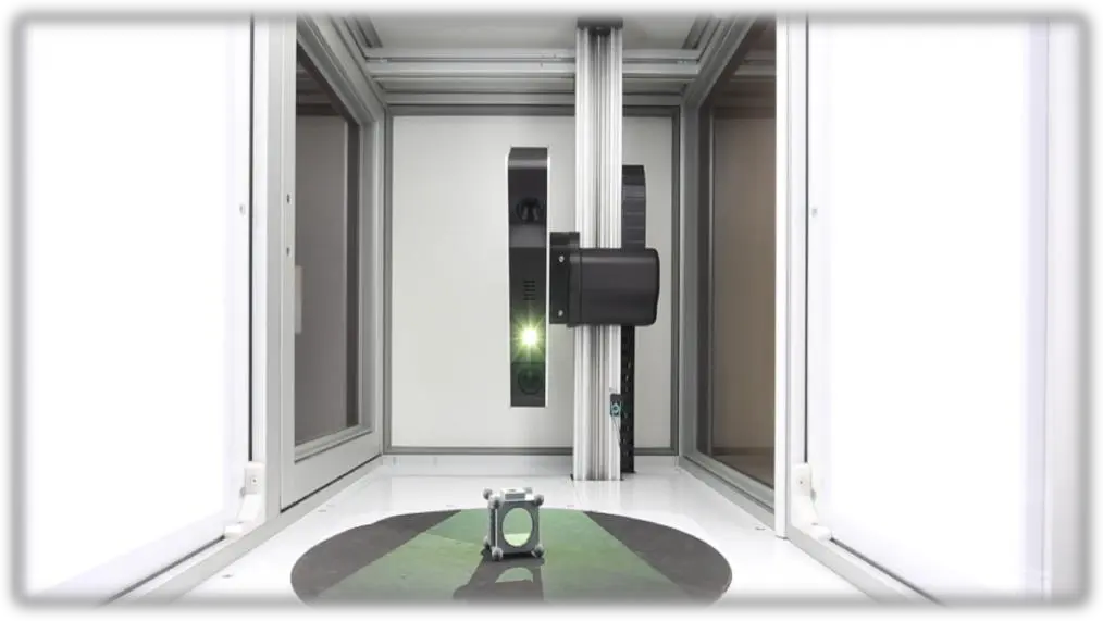
Interior of Taraz PM station with rotating table and optical system
Measuring area with component on the rotating table and acquisition from different orientations.

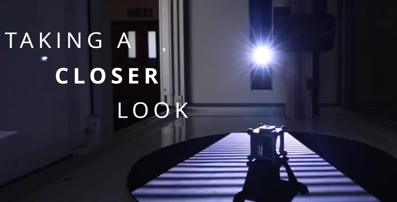
PROJECT FOR OFFICINE

An independent station for final inspection

The closed structure creates a controlled acquisition environment and brings optical metrology close to production. The configuration is optimized according to the piece and process, defining useful volume, sensors, movements, recipe and loading mode.

Quick SetupRepeatable acquisitionsDifferent materialsComplex shapesDedicated recipesCompliance Reports

APPLICATIONS

Where the single view is not enough

PM is indicated when the shape, orientation and completeness of the cover are decisive.

01

Processed components

Final control of shapes, quotas and deviations after milling, turning or grinding.

02

Additive production

Reconstruction of free-form components, curved surfaces and geometries produced by 3D printing.

03

Automotive and EV

Verification of components, supports, casings and assembly parts with respect to CAD.

04

Aerospace

Traceable inspection of complex parts and critical surfaces from multiple directions.

05

Assembly

Control of relative position of visible characteristics and overall geometry.

06

Research and pre-series

Quick comparison between real component, nominal model and subsequent process iterations.

PM O ASM?

Two complementary platforms

The choice depends on the result to be obtained, not only on the size of the sample.

CriteriaTaraz PMTaraz ASM
Main objectiveFull 3D shape and geometric complianceTopography, local form and aerospace roughness
AcquisitionMore views with traslation and tracked rotationAxial measurement from a prevailing orientation
TechnologyFrange projection + photogrammetryBlue light fringe projection
Ideal componentsComplex and three-dimensional geometryRelatively accessible or extended surfaces
ResultCast 3D model and CAD comparison2.5D point clouds and surface parameters

CONFIGURATION ON APPLICATION

Performance defined on the real component

Measurement range, accuracy, number of views, sensors and cycle time depend on size, geometry, material, finishing and tolerances required.The PM configuration is then defined after an application evaluation and sample test.

Technical deepening Taraz Metrology →

A COMPONENT TO REMEMBER?

We value coverage, references, tolerances and CAD comparison.

Request a PM test
← Back to products

METROLOGY LASER POINT AUTOFOCUS

Mitaka PF‑60

3D contactless profilemeter to measure shape, profile and roughness on extended areas, from mirror surfaces to transparent materials, without composition of fields.

Test PF‐60 on your sample
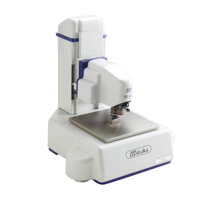
Laser profile Mitaka PF-60 original
60 × 60 mmMeasuring range XY
0.1 μmResolution of the X-Y stairs
0.01 μmResolution of the AF scale
87°Measurable maximum angle

MORE VELOCE, SEMPLICE AND PRECESS

Large submicrometric areas and details with the same size

The Scan AF function is up to 50 times faster than the previous Mitaka method.The laser probe with a range of 0.5 μm and the precision XY table directly measure areas of tens of millimeters up to the submicrometric scale.

The PF‐60 is suitable for deformations of precision components, molding defects, wear volume and quantitative evaluation of scratches and tribological surfaces.
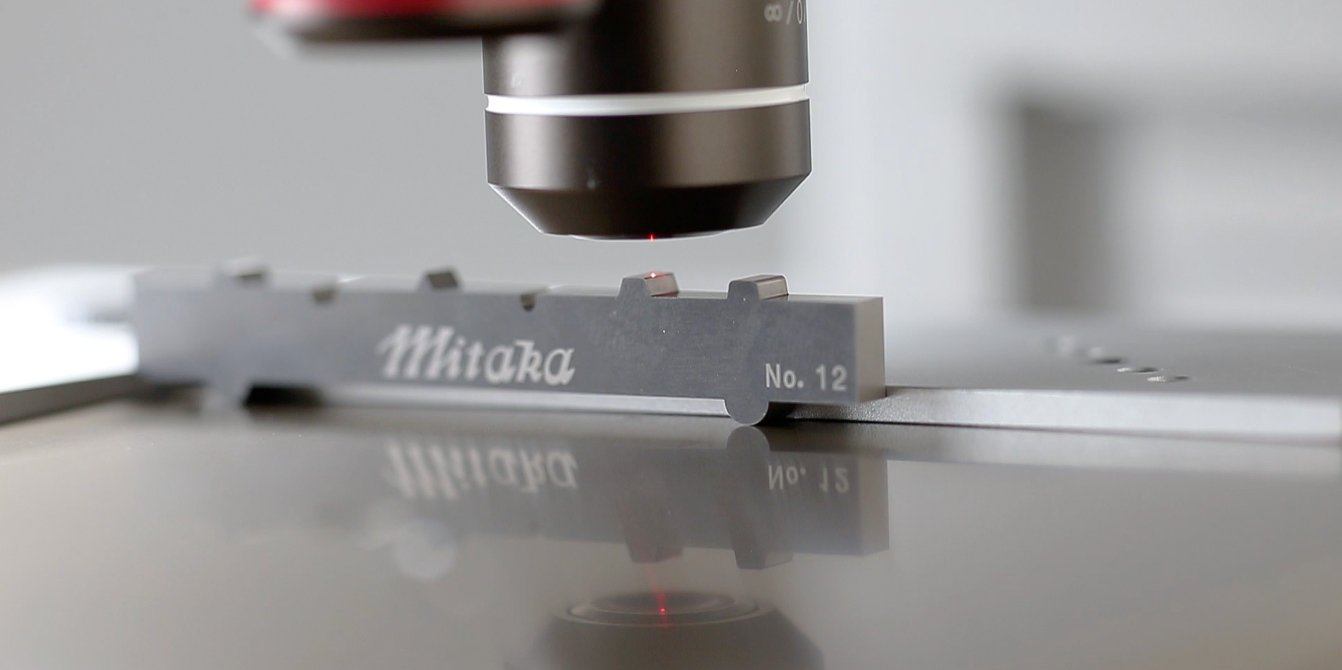
Contactless measurement with Mitaka PF-60

PRESTATIONS OF MEASURE

Quick 2D and 3D scanning, without stitching

The XY stage acquires coordinates over the entire race. The data comes from the continuous measurement of points and not from the union of separate images.
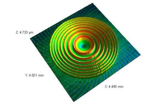
3D measurement of a Fresnel lens with PF-60

High precision extended area

Example of Fresnel lens: X 4,498 mm, Y 4,501 mm, Z 4,733 μm.
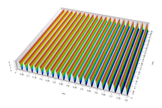
3D measurement of a lattice with PF-60

Quick AF scan

A measure requiring 1 hour and 20 minutes is performed in about 6 minutes; a 2D profile from 8,000 points takes about 15 seconds.
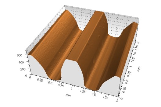
3D measurement of a microengineering with PF-60

In pursuit of strong slopes

The autofocus sensor detects low levels of reflected light and measures steps and inclined surfaces up to 87°.

MAIN POINT AUTOFOCUS

Fire becomes a co-ordination of share

The 635 nm laser, with a power of less than 1 mW, crosses the lens and forms on the surface an optical probe with a radius of 0.5 μm. The reflected light returns to the AF sensor, which detects in real time the motion of the spot and reports the microscope in the focus position.

The method is classified by ISO 25178‐6 and documented as ISO 25178‐605.

MEASURES AND ANALYSIS 3D

From shape to roughness, even on difficult materials

The reflectance of the surface affects little to the extent: the system operates on specular, transparent surfaces and with strong gradient variations.
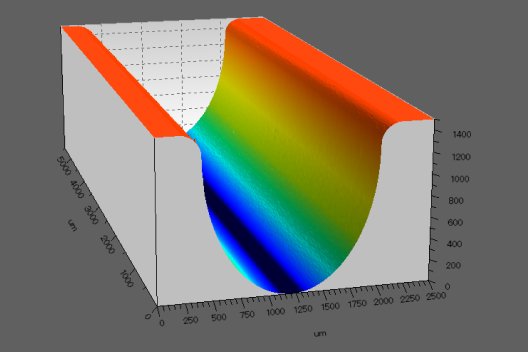
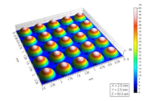
2D profile with resolution 0.01 μm3D shape and shape removalProfile extractionImage of the area and simple dimensional measuresRoughness and wear volumeDeformation and ripple

TECHNICAL DATA PF‐60

Compact laboratory configuration

Installation envelope400 × 400 mm
Tool weight31 kg
Field X × Y × Z × AF60 × 60 × 60 × 10 mm
X-Y scale resolution0.1 μm
AF scale resolution0.01 μm
Method of measurementFocus ISO 25178‐605
Lasers635 nm, <1 mW

Data and images translated from official page Mitaka Kohki.

DUE CONFIGURATIONS

Choose the system based on geometry

Mitaka PF-60EXTESE AREA

PF-60

Autofocus laser microscope with motorized XYZ board. Standard area 60 × 60 × 10 mm; continuous scanning without composition of visual fields.

  • Resolution XY 0.1 μm
  • Profile, shape and roughness measures
  • CSV/STL for CAD, FEM and reverse engineering
Mitaka MLP-3PSCANSION TO THE AROUND

5-axis MLP‐3P

Laser CMM with high-speed rotation and autofocus to measure internal, external surfaces and three-dimensional component profiles.

  • Pieces up to 120 × 120 × 120 mm
  • 5 programmable motorised axes
  • Contour, rotondity, gears and calibers

MITAKA APPLICATIONS

Where conventional optical measurement meets its limits

01

Thin films and wafers

Step height, texture, roughness and profiles on reflective, absorbent or transparent surfaces.

02

Optics, lenses and films

Shape, profile and roughness of transparent elements without contact with the surface.

03

Microcomponents and MEMS

Geometry, cavity and dislivelli with submicrometric laser spots and nanometric resolution.

04

Tools and edges

Ripid corners, rays, sharp profiles, wear and cutting characteristics.

05

Gears and cylindrical components

Profiles, rotondity, internal and external surfaces by scanning MLP‐3P all‐around.

06

Reverse engineering

STL/CSV export, CAD comparison, prototyping and FEM analysis data.

07

Additive manufacturing

Topography, roughness, geometric compliance and characterization of complex surfaces.

08

Production Metrology

Repeatable presets, automatic scans, reports and homogeneous batch control.

PRINCIPLE OF MEASURE

Point Autofocus Profiling

The laser spot is focused directly on the surface and the autofocus sensor follows the height in real time. The XY movement generates continuous three-dimensional coordinates, reducing the criticalities typical of the fire and optical stitching variation.

ISO 25178‐605Established non-contact method for measuring areal surface texture.No stitchingThe topography derives from contiguous lines of measured points, not from the union of images.Live measurementImages, parameters and results are available during the acquisition.

PORT US THE COMPONENT

We compare PF‐60 and MLP‐3P on your real geometry.

Request the Mitaka test
← Back to products

POLYRIX · SURROUND 3D SCANNERS

PolyScan X Series

Automatic, complete and secure 3D Metrology: a fixed network of cameras and projectors acquires the component from each direction, without robot trajectories and without risk of collision.

Evaluate a PolyScan application
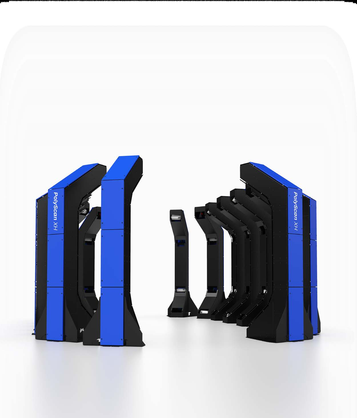
Polyrix PolyScan X Series
6-8 minScan time from XS to XL6
±0,010 mmXS Volumetric Accuracy
0.25-6,5+ mComponent Size
12-32+Cameras and projectors

_

Full cover without moving the scanner

PolyScan is a multi-sensor 3D measurement system without moving parts. Modular columns distribute cameras and projectors around the piece to acquire millions of points in a single cycle.
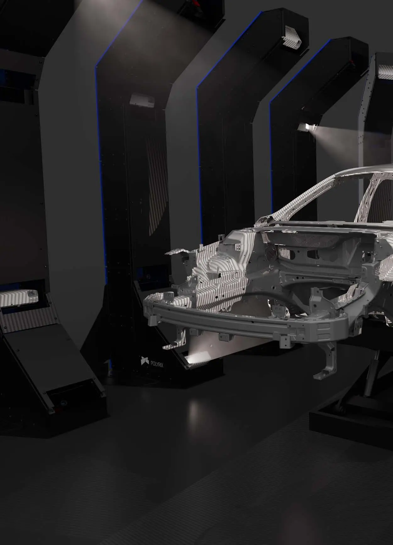
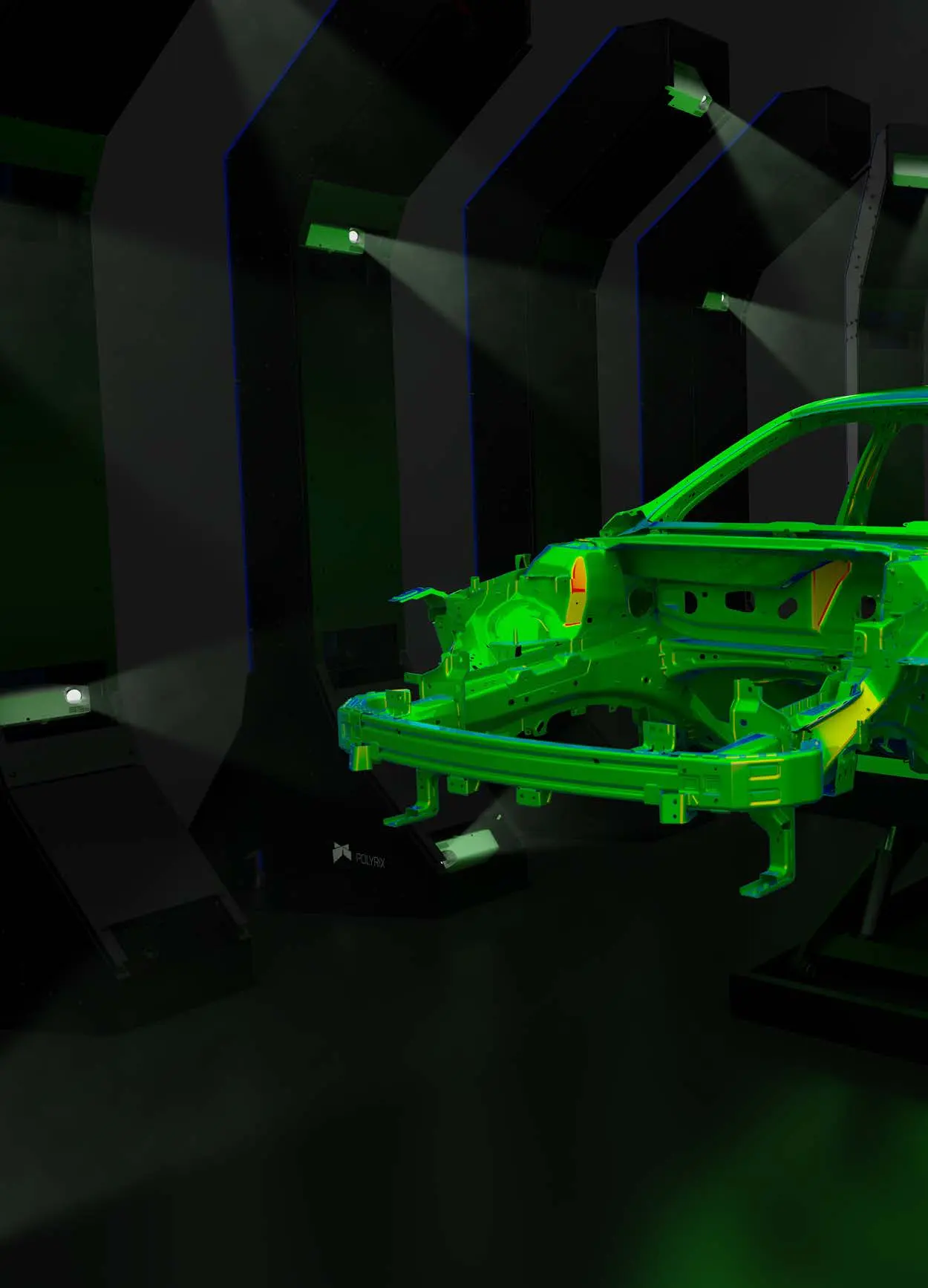
  • No trajectory programming
  • No typical safety requirement of moving robots
  • No complex recording with targets or software
  • Minimum processing and acquisition without operator intervention
Surround 3D Polyrix scanning of an automotive shell
Complete dimensional acquisition of an automotive structure.
Polyrix LiveInspection with color map projected on the component
LiveInspection: the deviation map is projected directly onto the piece.

LIVEINSPECTION

The result becomes immediately readable on the component

A matrix of high-definition projectors displays deviation color maps on the piece. The operator can change the map and receive dynamic and intuitive feedback during the evaluation.

3-4Quicker scan than manual or robotic scanners
Zero targetNo application and removal of references before and after scanning
FixedThe structure does not move: less complexity and maintenance
AutomaticAcquisition without human intervention and repeatable reports

_

From the small paddle to the large structure

Modular configurations with 6, 12 or 24 MP cameras, Full HD color projectors and 4K optional.
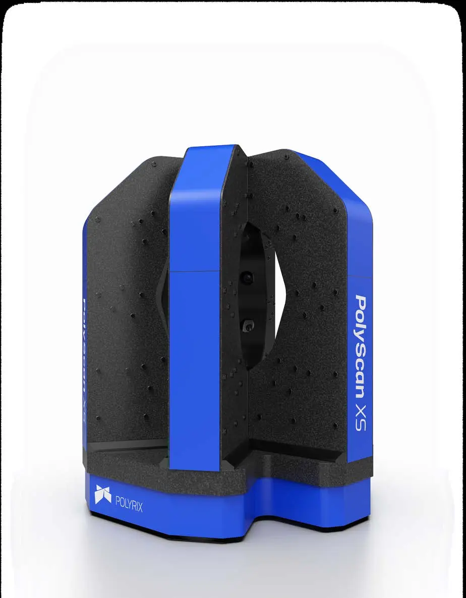
Polyrix PolyScan XS

XS

Piece
up to 0.25 m
Accuracy
±0,010 mm
Scanning
6-8 min
Sensors
12 + 12
Polyrix PolyScan XM

XM

Piece
up to 0.75 m
Accuracy
±0,020 mm
Scanning
6-8 min
Sensors
12 + 12
Polyrix PolyScan XL

XL6 / XL8 / XL10

Piece
1,7-2.5 m
Accuracy
±0,040-0,060 mm
Scanning
6-15 min
Sensors
12-20 + 12-20
Polyrix PolyScan XH

XH

Piece
up to 6.5+ m
Accuracy
±0,200 mm
Scanning
16-24+ min
Sensors
32+ + 32+

Accuracy based on third-party evaluation according to VDI/VDE 2634 part 2, accredited ISO 17025 laboratory and NIST traceability, as indicated in Polyrix documentation.

APPLICATIONS

Automatic control along the entire process

dimensional certification, in-process quality control, component inspection, moulds and matrices, repair assistance. Industries: automotive, aerospace, medical, defense, industry and energy.

Supported software: PolyWorks | Inspector plug-in, PolyScan Control Center and Polyrix API.
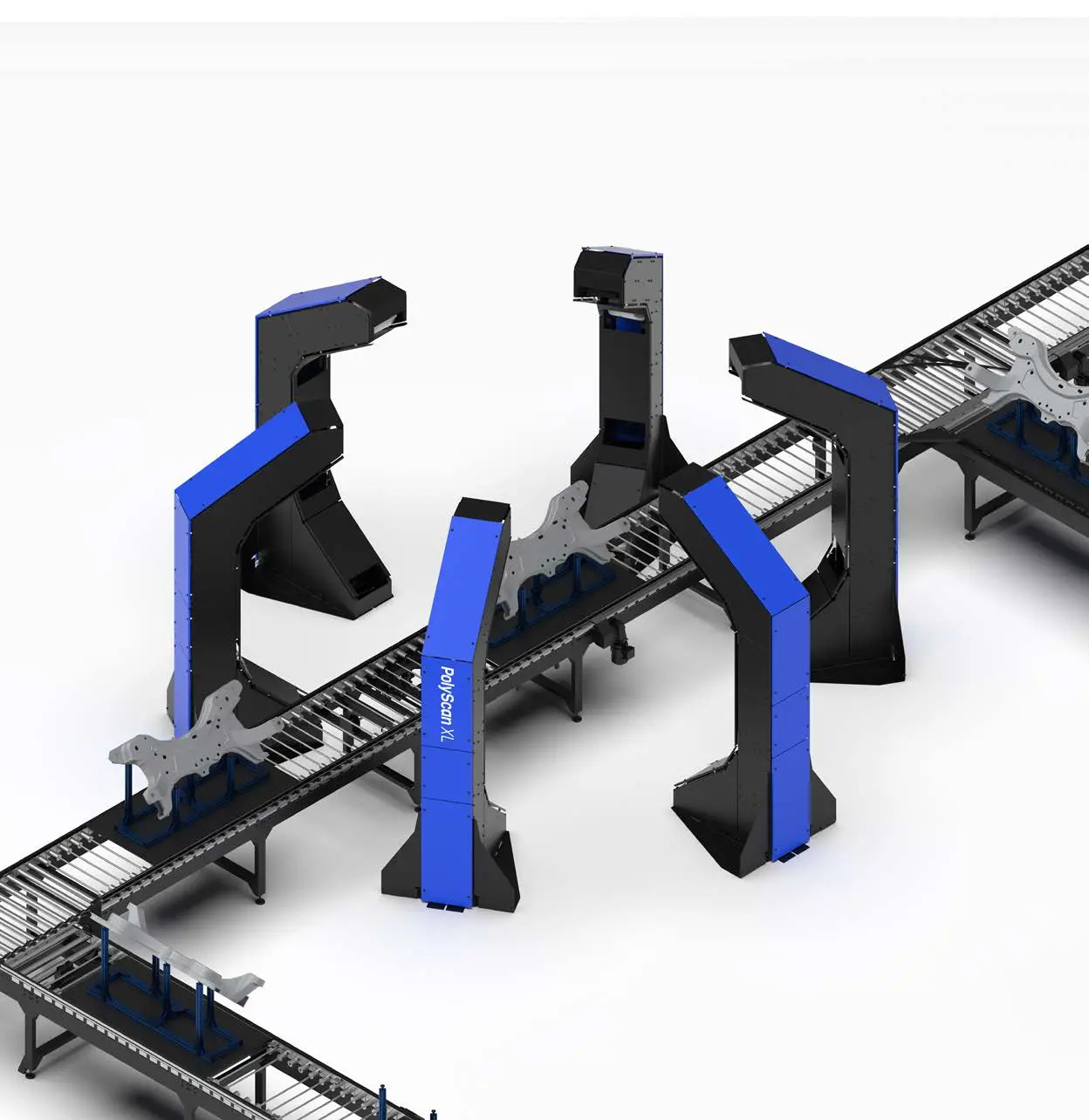
Polyrix PolyScan system integrated into an automated line

AUTOMATIC DIMENSION CONTROL

We define volume, accuracy and cycle time of your application.

Talk to a specialist
← Back to products

TOMOGRAPHY X-RAY FROM BANCO

nanoVoxel‑1000

Non-destructive three-dimensional analysis of the internal structures of small and medium samples, with compact laboratory configuration.

Evaluate your CT sample
nanoVoxel-1000
300 nmReliability of details
110 kVX‐ray sealed source
145 × 114 mmArea of the detector
15 kgMaximum sample weight

FEATURES

Compact micro‐CT, without dissecting the sample

Sealed source, flat panel detector from 2.940 × 2.304 pixels and samples indicating up to 300 mm in diameter and 200 mm in height.

Analysis of internal defects

Porosity, inclusions, cracks, assemblies and hidden geometries.

Imaging 4D in situ

Possible integration with mechanical or thermal load devices to observe the evolution of the sample.

Compact installation

Indicative dimensions 900 × 500 × 660 mm and vibration isolation system.

Application configuration

Hardware and software can be adapted to material, density and inspection target.

GUARDING DENTRO SENZA DISTRUGGIE

We value contrast, density and resolution on the sample.

Request a CT test
← Back to products

ST‐COBOT · EXPENDITURE AND AUTOMISED MEASURE

We see the invisible.
We measure the impossible.

3D digital microscopy, collaborative robotics and artificial intelligence for accurate, intelligent and non-destructive quality control, in the laboratory and in line of production.
AccuracyIntelligenceProductivity
HIROX Robotic Cell with Cobot for Automated Inspection and Measurement

ROBOTICA AND ARTIFICIAL INTELLIGENCE

From the position of the piece to the quality decision

We design control around the sample: handling, optical acquisition, measurement, classification and reporting.The performance occurs with an application test and agreed acceptance criteria.

01

Positioning

Cobot, portal or pick and place present the component in the defined poses.

02

Acquisition

HIROX, NPS and other sensors acquire images, profiles and surfaces in the control points.

03

Analysis

Methrological recipes and IA models identify candidate abnormalities and measure deviations.

04

Exit

Validated rules produce OK, verification or discard with images, measurements and traceability.

Robotics and IA for quality control — watch video
HIROX portal for programmed inspection of electronic cards
MICROELETTRO.

PCB and assemblies

Repeatable scans of tracks, welds, bonding and contamination, with localization of areas to be checked.

Robotic cell to present components to the optical system
MECHANICAL

Bearings and Components

Cell concept with draw from the line, presentation under the optics, measurement of geometries and surfaces and return of the outcome to the process.
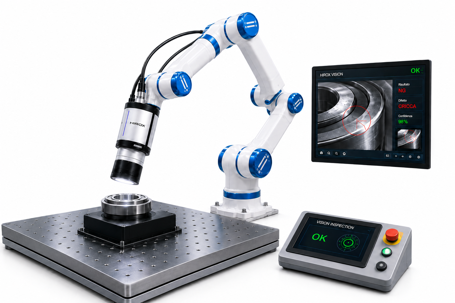
Cobot for inspection from multiple perspectives
COMPLESS HOURS

Multidirectional Inspection

The robot orientates optics or pieces to reach difficult edges, cavities and sides; the recipe records poses and acquisition parameters.

The images show configurations and application concepts. The possible IA classification requires representative data, validation and performance verification on the specific process.

ST‐COBOT

The microscope automatically reaches every control point

The six-degree robotic stave allows for high-precision encoded and programmable movements. The poses, sequences and capture parameters can be recalled to control components and batches in a repeatable way.

  • Programmable robotic handling on multiple sides of the component.
  • Possible translation unit on the robot's wrist for profile and roughness measurements.
  • Configuration prepared for online integration and data exchange with the factory.
ST-COBOT for automated inspection
6 degrees of freedomAutomatic control of surfaces, edges, cavities and distributed points.

CONTROLS IN LINE

Seven coordinate functions in one platform

From fault detection to data management: the cell can combine microscopy, metrology, automatic analysis and process traceability.

01

Micro Defect Detection

Automatic detection of micro-defects and abnormalities with artificial intelligence algorithms.

3D Map for Detection and Measurement of Microscrapers
02

Dimensional 3D scanning

High-resolution 3D acquisition for dimensional measurements and geometric verifications.

High-resolution three-dimensional reconstruction of a surface
03

Particle Analysis

Automatic analysis of particles, contamination and residues with AI-assisted classification.

Multi-spectral microscopic image for particle analysis and classification
04

Topography and roughness

3D measurement and analysis of surface topography, profiles and roughness parameters.

Three-dimensional chart of topography and surface roughness
05

Automation and IA

Intelligent and adaptive inspection to support real-time control decisions.

HIROX Cobot for Artificial Intelligence Automated Inspection
06

Tracking and Data Management

Management, storage and traceability of inspection data and process reports.

Automated cell with software interface for data management and traceability
07

Multitechnology Related Analysis

Integration of multiple technologies for a complete and multi-dimensional reading of quality control.

Hybrid system for analysis related to complementary measurement technologies

Structural controls with PAUT technology

The phased array ultrasound (PAUT) is a non-destructive control technique. The probe directs and focuses the beam at different depths and angles to detect and locate internal discontinuity in welds, pipes and metal components.

For an automated inspection, you can evaluate the integration of a PAUT probe, correcting the ultrasound indications with the position on the piece and with optical or dimensional reliefs.

Non-destructive controls and shared laboratories

Ekoscan documents the use of PAUT for axial and circumferential welds. ENEA carries out non-destructive ultrasound characterization activities and provides research infrastructure available to enterprises and scientific entities in the manner of individual laboratories.

DEMO INTERACTIVE · VISION + IA

Automatic detection of defects

Choose a sample and start the simulation: the recipe locates the anomalies, applies configurable thresholds and returns a traceable outcome.

Circuit observed under the microscope with candidate anomalies highlighted automatically
Analysis complete
Inspection recipe PCB‑BOND‑04Confidence threshold ≥ 90%
AUTOMATIC EXITReview required

Four areas exceed the thresholds set in the inspection recipe.

Anomaly candidates
4
Average confidence
94%
Analysing time
2.8 s
  1. 01 · Track discrepanciesConfidence 97%
  2. 02 · Surface residueConfidence 95%
  3. 03 · Uneven edgeConfidence 93%
  4. 04 · MicroscratchConfidence 91%

Demonstration simulation on predefined samples; it is not a test result. Classes, datasets, thresholds and acceptance criteria are validated on the component and process of the customer.

1 · Automatic acquisition2 · Localization IA3 · Classification4 · OK / verification / discard
01

Higher quality

Increased accuracy, repeatability and reliability of control.

02

Non-destructive control

Secure and contactless optical technologies with the component.

03

Operational efficiency

Faster processes, reduction of waste and increased productivity.

04

Artificial intelligence

Support for decisions through classification and automatic analysis.
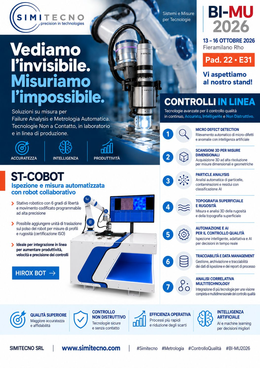
Locandina Simitecno BI-MU 2026 dedicated to online controls and ST-COBOT

BI‐MU 2026 · FIERAMILANO RHO

HiroxBot at BI‐MU 2026

From 13 to 16 October 2026, Simitecno presented solutions for Failure Analysis, automatic metrology and non-destructive quality control.

Select the inn to display it in the original size.

FROM THE MANUAL CONTROL TO THE AUTOMATIC RESEARCH

We define points, times, technologies and criteria of inspection.

Study a robotic cell

SAMPLE ANALYSIS

Tell us what you need to measure.

Send us the essential information: a Simitecno specialist will get back to you to assess the technology, the trial and the expected result.

  1. Describe the component, material or defect
  2. Specify the dimensions and the accuracy required
  3. We arrange a demo or a test on your sample
Prefer to talk right away?+39 06 7234320info@simitecno.it

Request an application assessment

This opens your email app with the details already filled in.

CONTACT

Speak directly with Simitecno’s technical team

For sales information, demonstrations, application analysis and support.

3D microscopy and metrology

IIT Milan Demo Centre

Center for Nano Science and Technology
Via Raffaele Rubattino, 81 · Milan

Book a demo →

A TECHNICAL ANSWER, NOT A GENERIC ONE

Tell us what you need to observe, measure or automate.

Describe your application
← All events

Upcoming workshop roadmap

3D microscopy for cultural heritage

Non-invasive surface documentation and before/after comparison of restoration work.

Applications and technical path

  • Identification of craquelure, waxing and lifting.
  • 3D reconstruction of the deformations of pictorial film.
  • Study of texture, color alterations and damaged areas.
  • High resolution images for documentation and comparison over time.

A comparison of laboratories, research and industry, with observations and measures on the samples. Dates, venue and modes of participation will be communicated with the workshop calendar.

Information: mario.brecciaroli@simitecno.it · +39 335 6252606

← All events

Upcoming workshop roadmap

Integrated HIROX system for ceramics

3D microscopy, structured light and artificial intelligence for surface inspection, from raw material to finished product.

Applications and technical path

  • High resolution analysis of microstructures and superficial defects.
  • 3D measurements of topography and complex geometries.
  • Detection and classification of defects with image analysis and AI.
  • Applications in research and development and quality control in production.

A comparison of laboratories, research and industry, with observations and measures on the samples. Dates, venue and modes of participation will be communicated with the workshop calendar.

Information: mario.brecciaroli@simitecno.it · +39 335 6252606

← All events

Upcoming workshop roadmap

HIROX and MITAKA for thin films

Multiscale imaging and laser profilometry for the characterisation of thin films and photovoltaic devices.
Applications and technical path

  • Observation HIROX with polarized and coaxial light.
  • Measurement of profiles and height variations with MITAKA PF-60.
  • Case study: Nb₂O₅ niobium oxide film on borosilicate glass.
  • In the illustrated case: nominal thickness 20 nm; measured height variation of about 19.95 nm.

A comparison of laboratories, research and industry, with observations and measures on the samples. Dates, venue and modes of participation will be communicated with the workshop calendar.

Information: mario.brecciaroli@simitecno.it · +39 335 6252606

← All events

Upcoming workshop roadmap

3D microscopy for metallography

From microstructures to failure analysis: observation, 2D/3D measurement and quantitative image analysis.
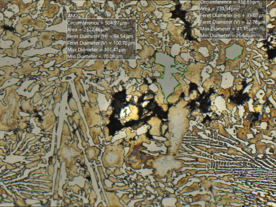
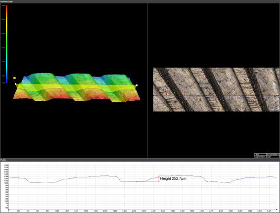
Applications and technical path

  • Analysis of fractures, microfractures, cracks and corrosion.
  • It has grains, studies of phases and inclusions.
  • 3D topography, roughness, porosity and process defects.
  • Workflow HIROX + MIPAR: segmentation, measurements, batches and reports with noted images.

A comparison of laboratories, research and industry, with observations and measures on the samples. Dates, venue and modes of participation will be communicated with the workshop calendar.

Information: mario.brecciaroli@simitecno.it · +39 335 6252606

Other application cases

Additional 3D scanning cases, counts grains, roughness and corrosion to compare microstructures and surface treatments.
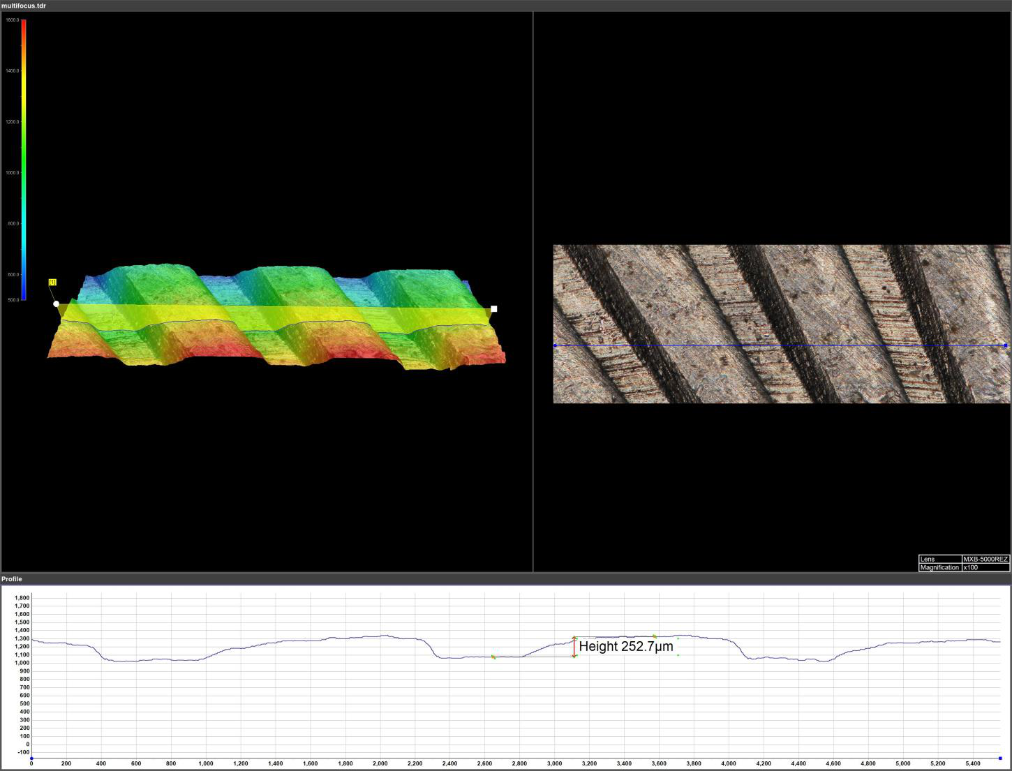

Download application cases
← All events

Upcoming workshop roadmap

3D digital microscopy for microelectronics

Inspection of assemblies, packaging and integrated circuits with 360° imaging, stitching and 2D/3D measurement.
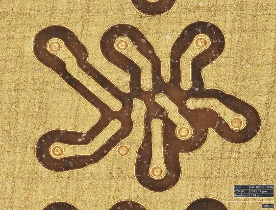
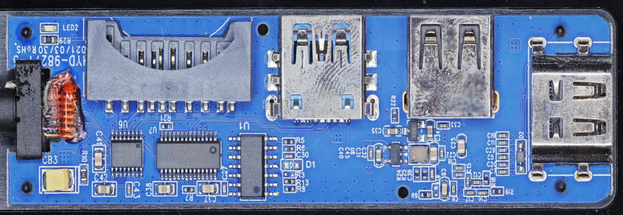
Applications and technical path

  • Control of welded components, joints and flux residues.
  • Observation of wafer pattern, tracks and details of microchip.
  • Search for microcracks and process defects on accessible surfaces.
  • Confocal profile NPS and 3D reconstructions for surface characterization.

A comparison of laboratories, research and industry, with observations and measures on the samples. Dates, venue and modes of participation will be communicated with the workshop calendar.

Information: mario.brecciaroli@simitecno.it · +39 335 6252606

← All events

Upcoming workshop roadmap

3D microscopy for fashion and jewellery

Fibres, leathers, finishes and jewellery: observing the details and documenting material quality.
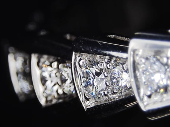
Applications and technical path

  • Morphology of natural and synthetic fibres: defects, breakages and contaminations.
  • Microfessure, finishing and wear of skins and leathers.
  • Quality of polishing, porosity and melting defects in precious metals.
  • Inspection of frames, frames and engravings; 2D/3D measurements.

A comparison of laboratories, research and industry, with observations and measures on the samples. Dates, venue and modes of participation will be communicated with the workshop calendar.

Videos and insights

Information: mario.brecciaroli@simitecno.it · +39 335 6252606

Other application cases

Application cases of 2D/3D measurements of defects, layer thicknesses and comparison of treated surfaces.
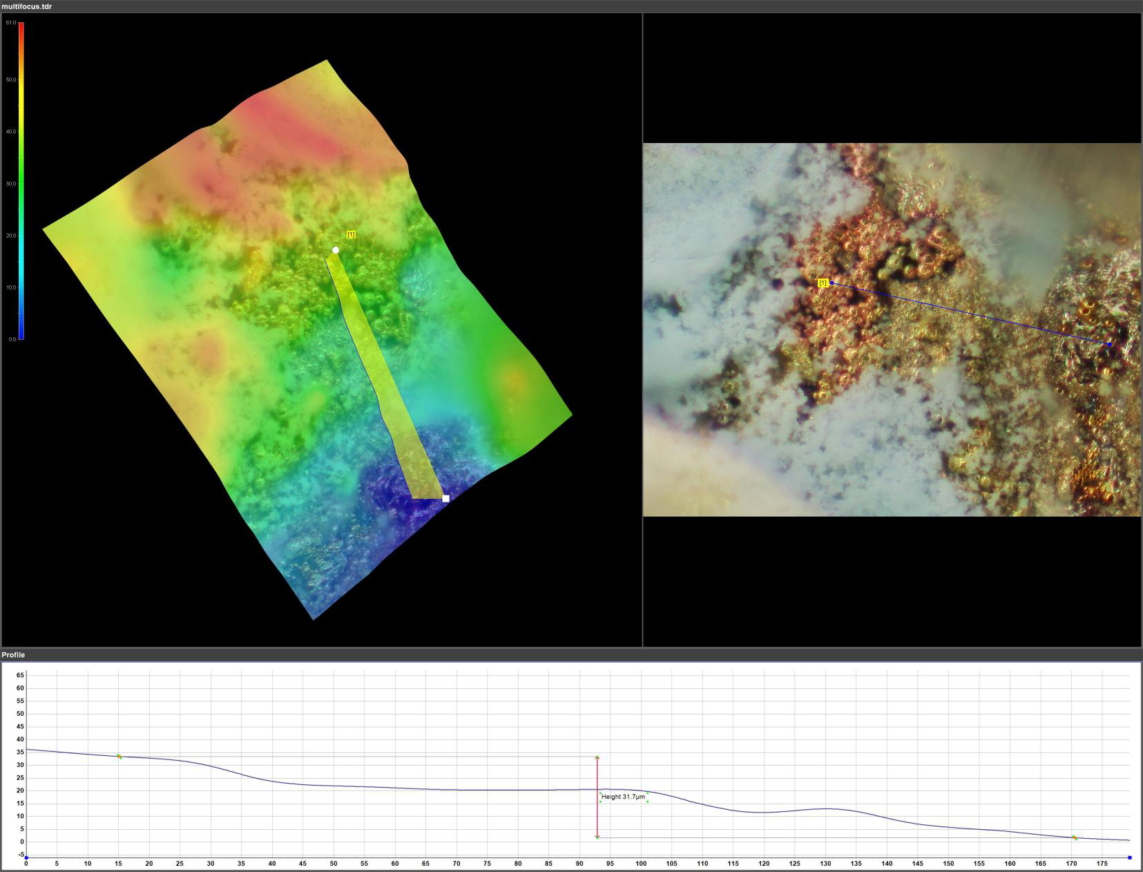
Download application cases
← All events

Upcoming workshop roadmap

3D microscopy for the life sciences

Zoology, entomology and botany through multifocal imaging, 3D reconstruction and sample documentation.
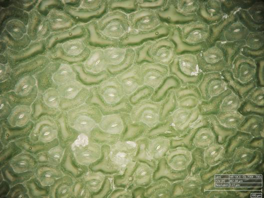
Applications and technical path

  • Study of scales, hairs, wings, composite eyes and other external structures.
  • Morphological comparison and character documentation for taxonomy.
  • Leaf surfaces, seeds, flowers and plant structures in 2D/3D.
  • Digital cataloguing of collections and production of images for research and teaching.

A comparison of laboratories, research and industry, with observations and measures on the samples. Dates, venue and modes of participation will be communicated with the workshop calendar.

Videos and insights

Information: mario.brecciaroli@simitecno.it · +39 335 6252606

← All events

Upcoming workshop roadmap

3D microscopy for Earth sciences

Rocks, minerals, fossils and sediments: morphological characterisation and microtopographic surveys.
Applications and technical path

  • Studio of microfractures, veins and superficial alterations of the rocks.
  • Analysis of crystals, inclusions and surface structures of minerals.
  • Documentation of fossils, fingerprints and microfossil remains.
  • Weaving, granulometry and porosity of sediments; introduction to the LIBS module for elementary analysis.

A comparison of laboratories, research and industry, with observations and measures on the samples. Dates, venue and modes of participation will be communicated with the workshop calendar.

Videos and insights

Information: mario.brecciaroli@simitecno.it · +39 335 6252606

← All events

Upcoming workshop roadmap

3D microscopy for forensic science

Micro-traces, exhibits and materials: imaging and measurement to document and compare observable details.
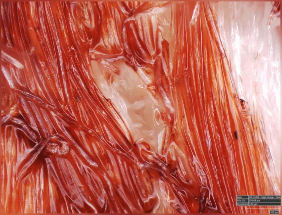
Applications and technical path

  • Morphological documentation of fibers, hair, glass fragments and paint.
  • Comparative analysis of surface traces and 2D/3D measurements.
  • Study of fractures, wear, corrosion and contamination of the finds.
  • Comparison of brush strokes and pattern processing, with image storage.

A comparison of laboratories, research and industry, with observations and measures on the samples. Dates, venue and modes of participation will be communicated with the workshop calendar.

Videos and insights

Information: mario.brecciaroli@simitecno.it · +39 335 6252606

← All events

Upcoming workshop roadmap

HIROX for biomaterials and scaffolds

From polymers to scaffolds: morphological analysis, topography and surface inspection for biomaterials research.
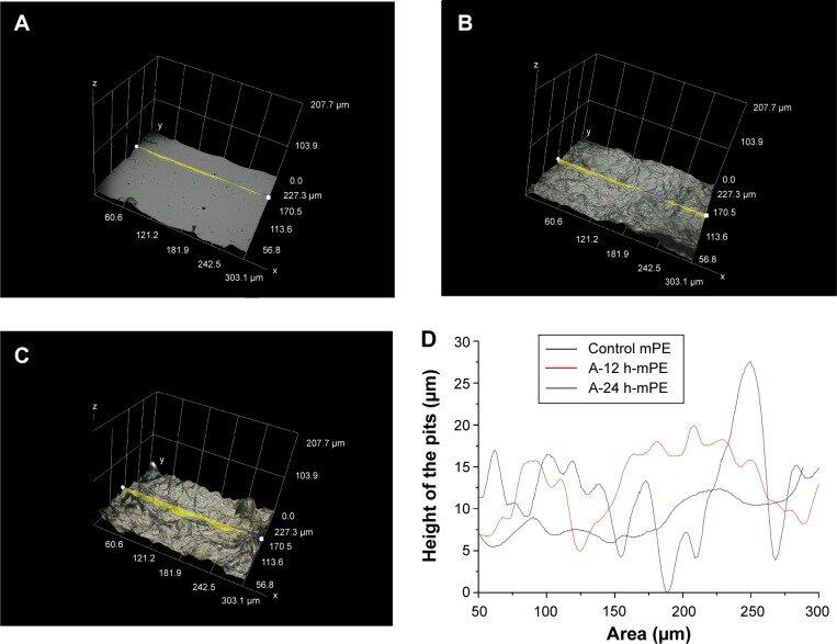
Applications and technical path

  • Morphology, roughness and superficial porosity of biomaterials and scaffold.
  • Inspection of surfaces after CNC machining or plasma treatments.
  • Biocompatible ceramics: observation of fractures and microfractures.
  • Illustrated case studies: PDMS films with NdFeB particles and mPE surfaces functionalized with natural extracts.

A comparison of laboratories, research and industry, with observations and measures on the samples. Dates, venue and modes of participation will be communicated with the workshop calendar.

Videos and insights

Information: mario.brecciaroli@simitecno.it · +39 335 6252606

← All events

Upcoming workshop roadmap

3D microscopy for glass and coatings

Glass failure analysis, surface defects and coatings: illumination, dimensional measurement and topography.
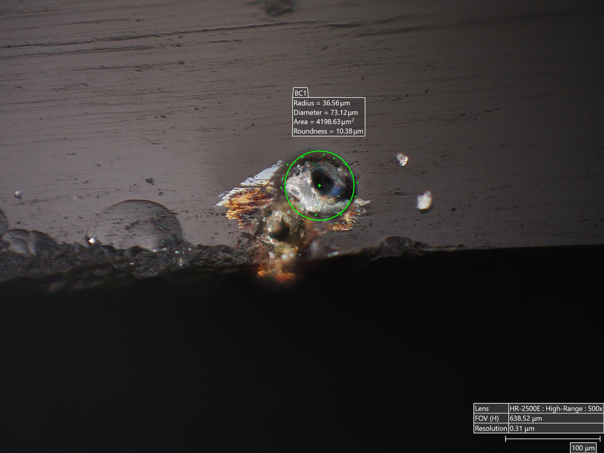
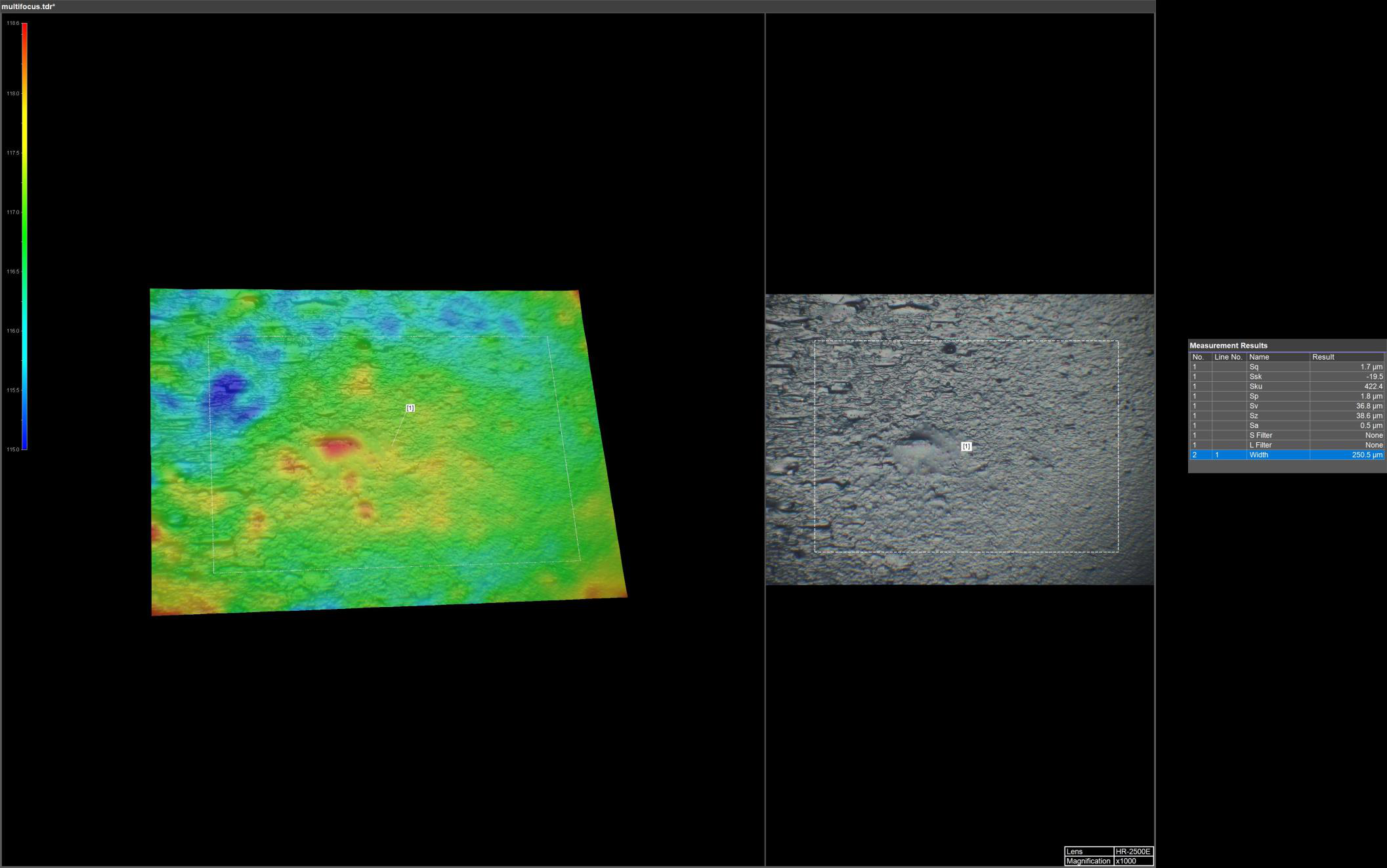
Applications and technical path

  • Comparison of fracture surfaces with epi-coaxial and rotating optics.
  • Observation of defects with different lighting modes.
  • 2D sizes of defects, 3D textures and roughness of surfaces.
  • Study of bubbles, cracks, delaminations and uniformity of coatings.

A comparison of laboratories, research and industry, with observations and measures on the samples. Dates, venue and modes of participation will be communicated with the workshop calendar.

Videos and insights

Information: mario.brecciaroli@simitecno.it · +39 335 6252606

← All events

Upcoming workshop roadmap

3D microscopy for fibres and textiles

From single fibres to fabric: measurement, stitching and documentation for laboratory, development and quality control.
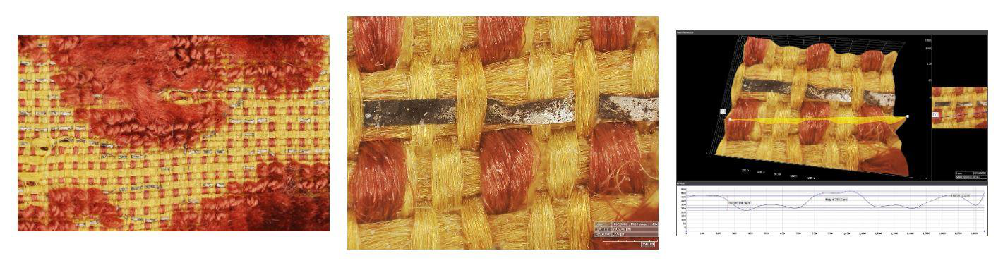
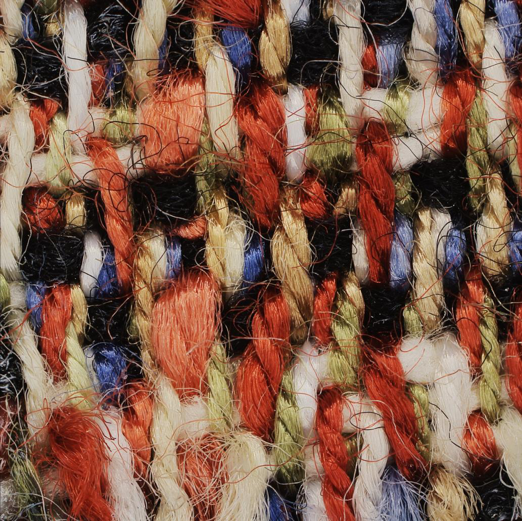
Applications and technical path

  • morphological analysis of natural, synthetic and mixed fibres.
  • Study of weaving, uniformity and density of weaving and warp threads.
  • Evaluation of finishes and treatments; identification of irregularities.
  • 3D reconstructions, prototyping and traceability of analysis.

A comparison of laboratories, research and industry, with observations and measures on the samples. Dates, venue and modes of participation will be communicated with the workshop calendar.

Videos and insights

Information: mario.brecciaroli@simitecno.it · +39 335 6252606

Other application cases

Stitching of extended areas to document the structure of the sample and compare different areas.
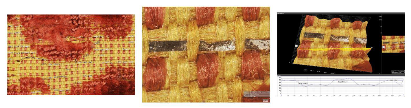
Download application cases
← All events

Upcoming workshop roadmap

Surface and failure analysis

From topography to defects: optical metrology, coatings, wear and inspection automation.
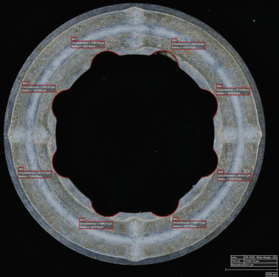
Applications and technical path

  • Roughness, texture, fractures and cutting edges.
  • Measures not in contact with depth, heights, angles and distances.
  • Uniformity of coatings and comparison before/after treatment.
  • HiroxBot, image analysis and traceability for quality control workflows.

A comparison of laboratories, research and industry, with observations and measures on the samples. Dates, venue and modes of participation will be communicated with the workshop calendar.

Videos and insights

Information: mario.brecciaroli@simitecno.it · +39 335 6252606

← All events

Upcoming workshop roadmap

3D microscopy for paper, board and packaging

Fibre structure, finishes and defects for the development and inspection of packaging materials.
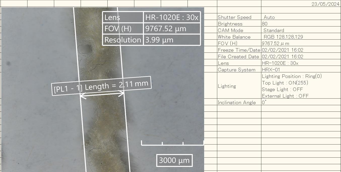
Applications and technical path

  • Observation of the disposition and integrity of fibers.
  • Study of surfaces, coatings and laminations.
  • Identification of microfractures, inclusions and irregularities.
  • 2D/3D measurements and reports to compare samples and production processes.

A comparison of laboratories, research and industry, with observations and measures on the samples. Dates, venue and modes of participation will be communicated with the workshop calendar.

Videos and insights

Information: mario.brecciaroli@simitecno.it · +39 335 6252606

← All events

Upcoming workshop roadmap

Paper, printing and inks

Paper texture, ink distribution and print detail with dedicated imaging and illumination.
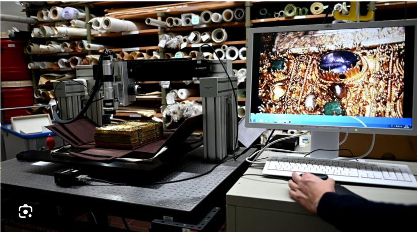
Applications and technical path

  • Study of fibers, pigments and ink distribution uniformity.
  • Stitching and 3D reconstructions on extended areas.
  • Comparison between observations in visible and infrared light.
  • Cases illustrated in PDF: papyrus, underlying designs and print details overlapped with a signature.

A comparison of laboratories, research and industry, with observations and measures on the samples. Dates, venue and modes of participation will be communicated with the workshop calendar.

Videos and insights

Information: mario.brecciaroli@simitecno.it · +39 335 6252606

← All events

Upcoming workshop roadmap

3D microscopy for composites

Fibres, matrix and laminates: multiscale inspection, dimensional measurement and defect analysis.
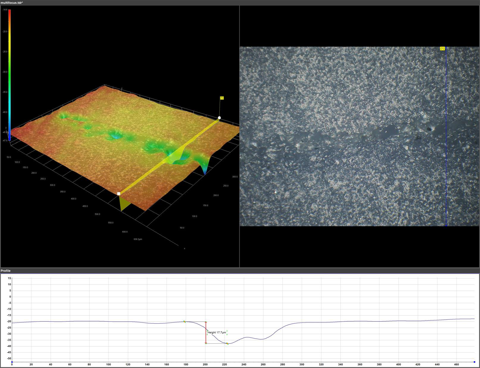
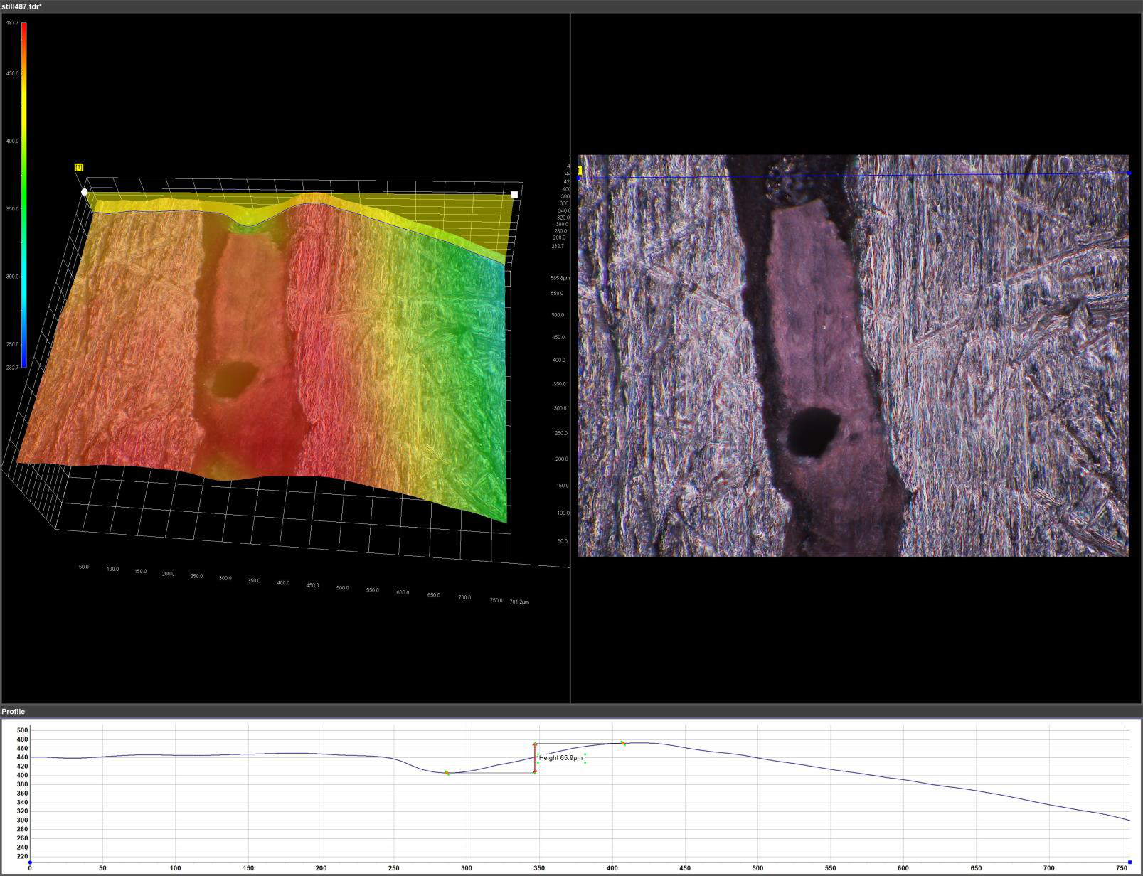
Applications and technical path

  • Areas rich in resin, fiber alignment and layer arrangement.
  • Measurements of the thickness of the layers and study of visible voids.
  • Stitching to extend the analyzed area and compare the laminate areas.
  • 3D measurement cases of defects and aerospace components inspection.

A comparison of laboratories, research and industry, with observations and measures on the samples. Dates, venue and modes of participation will be communicated with the workshop calendar.

Videos and insights

Information: mario.brecciaroli@simitecno.it · +39 335 6252606

← All events

Upcoming workshop roadmap

Dermatology, trichology, cosmetics and pharmaceuticals

Imaging and measurement for research, surface characterisation and quality control of products and materials.
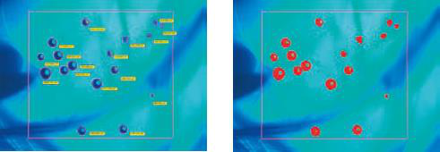
Applications and technical path

  • Observation of superficial morphology of hair and skin samples.
  • Nail polish test and make-up; characterization of surfaces and packaging.
  • Particle count through binaryization and RGB selection.
  • Image documentation and results for research and quality control activities.

A comparison of laboratories, research and industry, with observations and measures on the samples. Dates, venue and modes of participation will be communicated with the workshop calendar.

Videos and insights

Information: mario.brecciaroli@simitecno.it · +39 335 6252606

← Past Events

TRADE FAIR · LIVE TESTING

BI-MU – Milan

13–16 October 2026 · Fieramilano Rho · Pavilion 22 · Stand E31

System for BI-MU – Milan

Announced configuration

  • 1 gantry with Hirox
  • 2 Hirox with benchtop stands, one of them with NPS
  • 1 cobot with Taraz
  • 1 cobot cell with Hirox

Program and configuration announced for this appointment. Contact SIMITECNO to request materials or arrange a new application test.

mario.brecciaroli@simitecno.it · +39 335 6252606

Digital and confocal microscopy 3D and textured light

Advanced techniques for three-dimensional characterization of surfaces.The BI-MU program provided application analysis on samples with the Simitecno specialists.
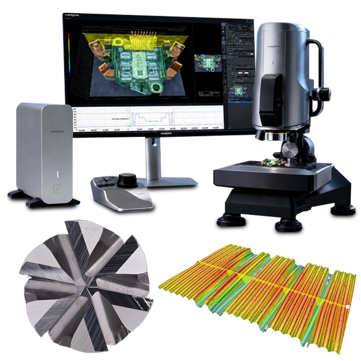
← All events

WORKSHOP

IIT workshop – Milan

Dates to be defined · IIT · CNST · Milan

System for the IIT Workshop – Milan

Exposed instrumentation

  • Hirox with benchtop stand
  • NPS

Contact the SIMITECNO team to agree to participation, demonstrations and samples of interest.

mario.brecciaroli@simitecno.it · +39 335 6252606

← Past Events

TRADE FAIR · APPLICATION DEMOS

A&T – Vicenza

27–29 October 2026 · Vicenza

System for A&T – Vicenza

Announced configuration

  • 2 Hirox with benchtop stands, one of them with NPS
  • 1 cobot with Taraz

Program and configuration announced for this appointment. Contact SIMITECNO to request materials or arrange a new application test.

mario.brecciaroli@simitecno.it · +39 335 6252606

← All events

WORKSHOP

IIT workshop – Milan

Dates to be defined · IIT · CNST · Milan

System for the IIT Workshop – Milan

Exposed instrumentation

  • Hirox with benchtop stand
  • NPS

Contact the SIMITECNO team to agree to participation, demonstrations and samples of interest.

mario.brecciaroli@simitecno.it · +39 335 6252606

← Past Events

WORKSHOP

CNR ISM workshop – Frascati

09 October 2026 · Frascati

System for the CNR ISM Frascati Workshop

Announced configuration

  • 1 Mitaka
  • 1 Hirox with Rvomax + NPS

Program and configuration announced for this appointment. Contact SIMITECNO to request materials or arrange a new application test.

mario.brecciaroli@simitecno.it · +39 335 6252606

← All events

OPEN HOUSE

Open House Werth Italia

24 and 25 November 2026 · 09:00–13:00 and 14:00–17:00 on both days

Werth Italia Srl · Via Sempione 17 · 21011 Casorate Sempione (VA)

Programme

Live demonstrations of Hirox 3D digital microscopy, multisensor CMMs and computed tomography.

Open access to Werth’s laboratories and discussions with experts on equipment, technologies and software for 3D optical measurement, multisensor metrology and computed tomography.

info@werth-italia.it · +39 0331 770746

SIMITECNO contacts

← All events

WORKSHOP

Automated microdefect detection and 3D surface topography

IIT workshop – Milan

Dates to be defined · IIT · CNST · Milan

System for the IIT Workshop – Milan

Exposed instrumentation

  • Hirox with benchtop stand
  • NPS

Contact the SIMITECNO team to agree to participation, demonstrations and samples of interest.

mario.brecciaroli@simitecno.it · +39 335 6252606

← All events

WORKSHOP

University of Messina workshop

Dates to be defined · University of Messina

System for the University of Messina Workshop

Exposed instrumentation

  • Hirox
  • Mitaka

Contact the SIMITECNO team to agree to participation, demonstrations and samples of interest.

mario.brecciaroli@simitecno.it · +39 335 6252606

← All events

TRADE FAIR

MECSPE

3–5 March 2027 · Location to be defined

System for MECSPE

Configuration to be confirmed

Contact the SIMITECNO team to agree to participation, demonstrations and samples of interest.

mario.brecciaroli@simitecno.it · +39 335 6252606

Technologies and applications

Illustrative images of SIMITECNO application themes. The MECSPE exhibition configuration is to be defined.
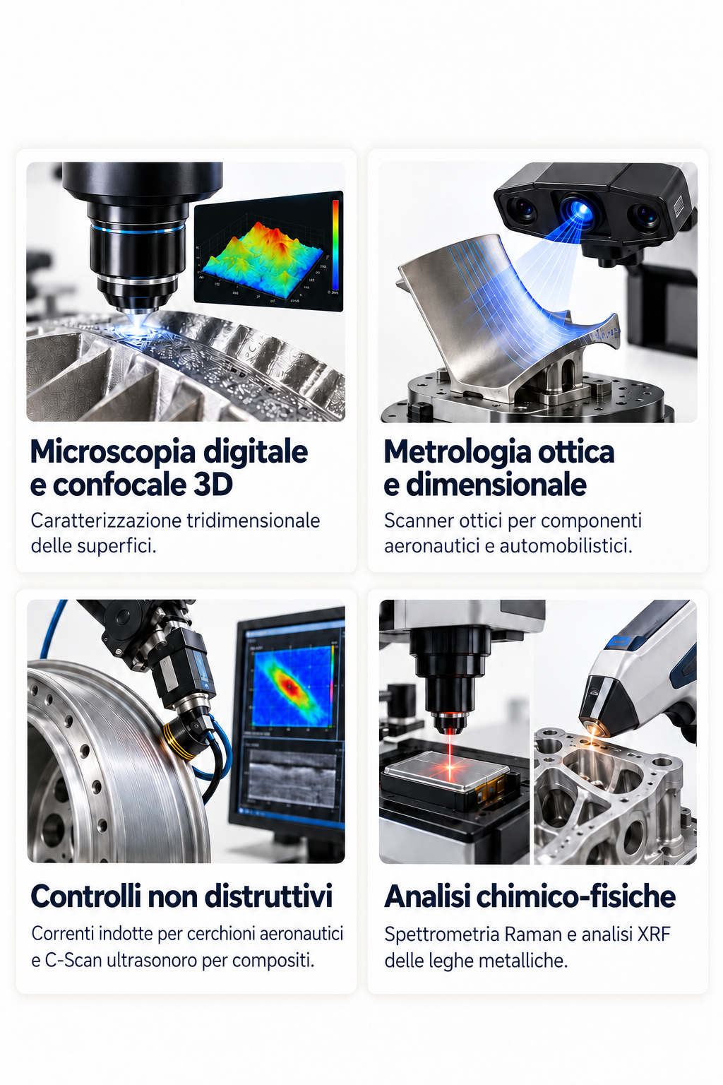
INTELLIGENCE

News

Technologies and applications for analysis and quality control of materials.

TECHNOLOGICAL FOCUS · 6 OCTOBER 2026

LIBS and HIROX: from image to elementary analysis

The microscopic observation identifies the point of interest.The LIBS spectroscopy helps to characterize its elementary composition.
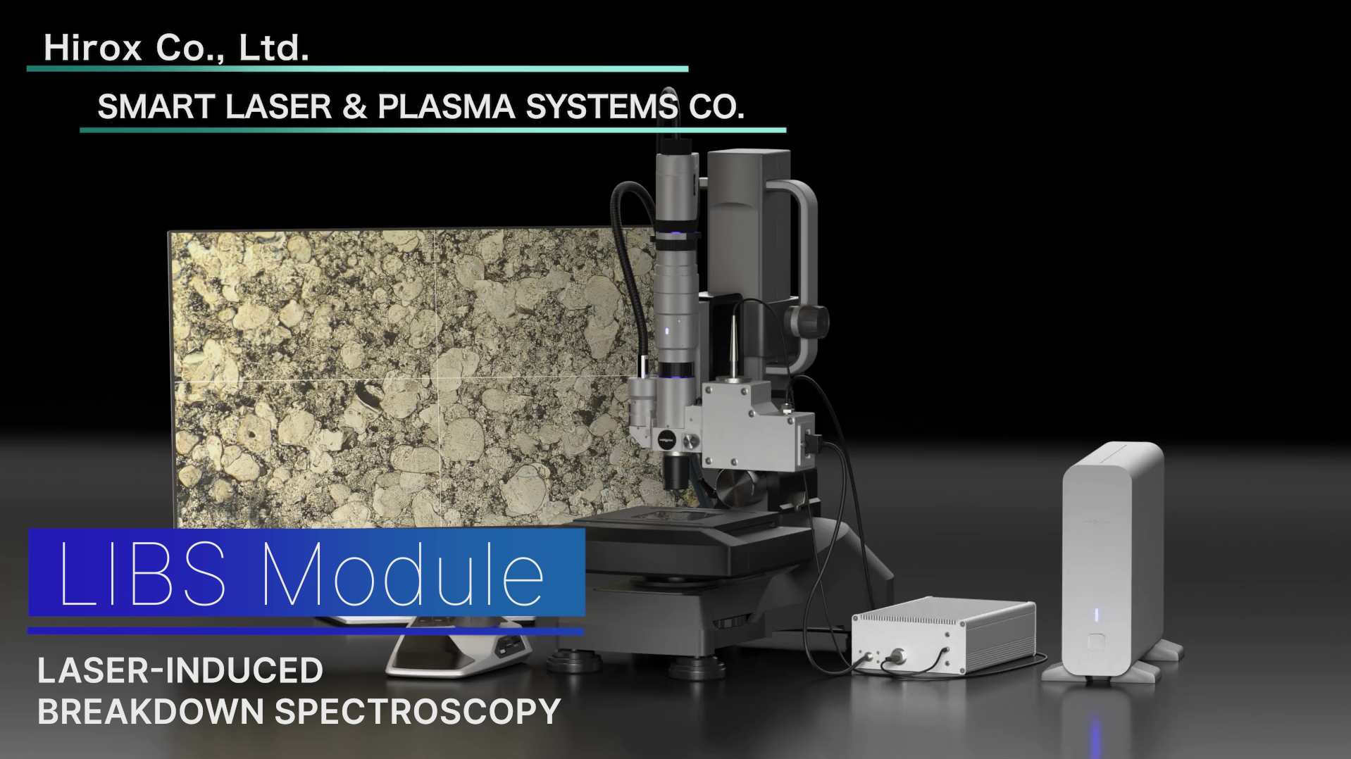
LIBS module combined with HIROX microscope, from promotional presentation
Configuration shown in HIROX / Smart Laser & Plasma Systems Co promotional material.

What is LIBS

LIBS means Laser-Induced Breakdown Spectroscopy, laser-induced plasma emission spectroscopy. A focused laser pulse removes a very small amount of material and generates a plasma.

The technique involves a local microablation: on the analyzed point a small crater can remain.

The value of matching with the microscope

The images of the presentation show a LIBS module combined with HIROX: sample observation, selection of an area and acquisition of the spectrum can thus be part of a related characterization path.

The correlation between morphology and composition can support the analysis of inclusions, particles and unevenness of materials.
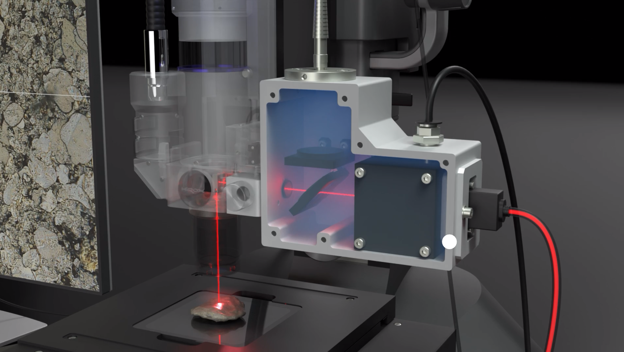
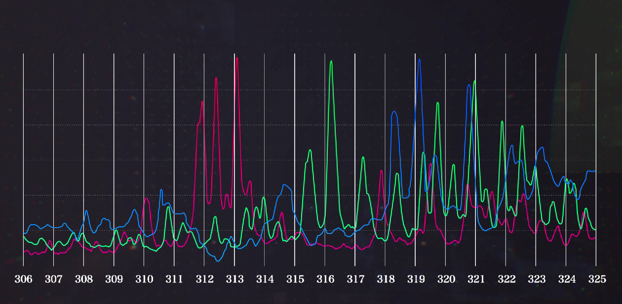
Applications of interest

  • Local characterization of metals and alloys.
  • Study of inclusions and foreign particles.
  • Analysis of ceramic materials, glass and geological samples.
  • Comparison between sample areas with different morphology.

From the demonstration to your sample

The detectable elements, detection limits and quantitative analysis possibilities depend on the material, configuration and calibration.

To learn more about HIROX–LIBS, share the material and area you want to analyze with SIMITECNO.

Request information about LIBS

TECHNICAL DOCUMENTATION · HIROX

MS-LIBS: microscope and elementary analysis in the same system

The Fujikin documentation presented at JASIS 2026 indicates the technical collaboration of HIROX and Smart Laser & Plasma Systems. The system focuses a pulsed laser on the point observed under the microscope and analyzes the light of the generated plasma.
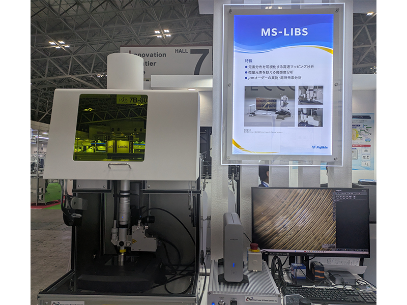
From the point observed to the composition

The brochure describes local analysis, two-dimensional mapping of the elements and deep analysis. The spot declared from 30 μm; the sensitivity is indicated in the ppm–% range, with performance to be evaluated on the material and the measurement conditions.

Comment application SIMITECNO. Correlating image and composition can help investigate a foreign particle, inclusion or uneven area. The mapping allows you to evaluate how the elements are distributed in the analyzed area.

Materials and applications

  • Metals and alloys: identification and study of segregation.
  • Materials for batteries: distribution and uniformity of elements.
  • Electronic materials: microcontaminants and localized analysis.
  • Deep analysis: variations of the composition from the surface to the inside.

The laser removes a small amount of material: it is necessary to evaluate the acceptability of the microablation on the sample.

RESEARCH · COMMENTARY BIBLIOGRAPHY

Publications: HIROX in LIBS research

These studies cite HIROX microscopes for the observation or measurement of samples in LIBS research. They document a complementary use of the microscope; they do not constitute a validation of the new MS-LIBS integrated system.

2025 · ACS OMEGA · MINERAL

Mixture Detection Using Deep-UV Raman-LIBS Autofocus-Based Compact Chemical Spectroscopic Sensor

HIROX RH-8800, 4000×. In Figure 4, microscopic images show the mineral samples W30, olivine and their mixture.

Comment SIMITECNO. Documenting morphology and distribution of grains helps to interpret the spectroscopic analysis of heterogeneous materials.

Read the publication · DOI 10.1021/acsomega.5c03801

2021 · APPLIED OPTICS · ADDITIONAL MANIFATTURE

Surface hardness determination of 3D printed parts using laser-induced breakdown spectroscopy

Jinwei Yang, Linghua Kong, Guofu Lian and Tongfei You · Applied Optics 60, 499–504. HIROX KH-1300 The study examines the relationship between LIBS signals and the surface hardness of the 18Ni300 maraging steel printed in 3D.

Comment SIMITECNO. Measuring the crater allows to document the effect of the laser on the surface and to support a geometrical control of the spectral response.

Read the publication · Applied Optics

2013 · SPIE · CULTURAL GOODS

Use of the LIBS method in oil paintings examination...

The work of E. Modzelewska and collaborators concerns the examination of oil paintings through LIBS and cites microscopes HIROX 8700 in the observation path.

Comment SIMITECNO. The microscopic observation can be accompanied by the study of painting surfaces and the documentation of the area analyzed.

Read the publication · DOI 10.1117/12.2020694

The SIMITECNO comments are interpretations and not quotes of the authors. Full texts and figures can be consulted with publishers according to the respective conditions of access. Sources consulted on 6 October 2026.

Initial images: presentation "screenshot promotional video" provided at SIMITECNO. MS-LIBS photos and brochures: Fujikin, with technical collaboration declared HIROX / Smart Laser & Plasma Systems.

_

Taking a closer look at materials for over 40 years.

We help industry and research to observe, measure and understand materials. We start from a sample, a defect or a concrete question and build the most suitable control path.
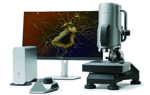
Simitecno technologies for microscopy and quality control of materials

OUR IDENTITY

Application expertise, tools and people.

Simitecno S.r.l. is based in Rome and operates in quality control of materials. We support laboratories, research centers and enterprises in the choice and integration of technologies for surface analysis, metrology and non-destructive controls.

Our work combines technical advice, sample demonstrations, system configuration, training and assistance.

HOW TO WORK

From problem to decision.

01

Let's hear the case

Material, geometry, defect and measurement target define technical demand.

02

Let's try on the sample

We compare technologies and configurations on a real case, in the laboratory or at the customer.

03

We integrate the solution

From the bench to the automated cell, we adapt data acquisition, measurement and flow to the process.

04

We stand by the team

Training, application support and assistance help make control repeatable.

Demo Center Simitecno at IIT CNST in Milan

DIMOSTRATIONS AND RESEARCH

The test on your samples is the starting point.

In the Demo Center at IIT–CNST in Milan, you can explore the HIROX 3D digital microscope and the NPS confocal profilemetry. We work with the research world and with technological partners to translate new measurement capabilities into industrial applications.

Organize a demonstration →

THE COMPETENCES

A single interlocutor, complementary technologies.

3D digital microscopyOptical metrology and profilemetryNon-destructive controlsChemical-physical analysisRobotics and Artificial IntelligenceAutomation of quality control

We follow applications in aerospace, automotive, energy, microelectronics, manufacturing, cultural heritage and materials research.

WORK

What problem do you want to solve?

Tell us what you need to observe or measure: we will evaluate with you an application path and a test on the sample.

Contact Simitecno
← Back to the HIROX section

SCIENTIFICATION RESEARCH · BIBLIOGRAPHY

HIROX in research

A collection of publications documenting the use of HIROX digital microscopes in scientific studies. Each card indicates the application observed and refers to the original article, with figures and methodological details.

The opening photographs of the cards are illustrative images already present on the Simitecno website: they are not figures from the articles. The original scientific images are found in the related publications. The collection is selective and up-to-date, not an exhaustive census.

Do you have an application to check?

We can evaluate 2D/3D observations, topography and documentation of your sample with the most suitable configuration.

Request an application test
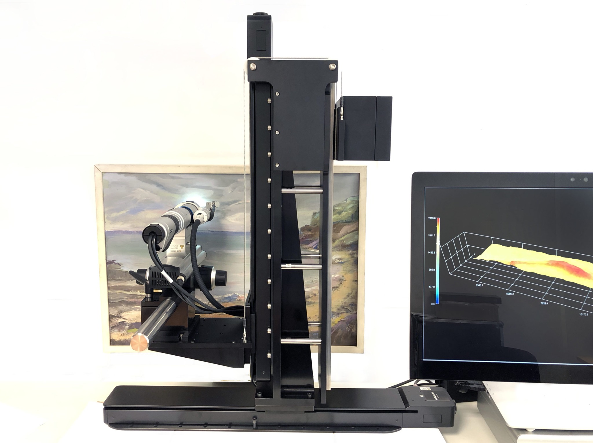
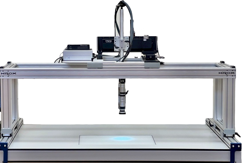
Analyse your sample

Search the site